Study: SLSC Photoacoustic Imaging to Reduce Skin Tone Bias
Traditional PA imaging struggles to read dark skin tones. A recent study says short-lag spatial coherence beamforming could help.

Traditional medical imaging technology has long struggled to obtain clear pictures of patients with Fitzpatrick skin phototypes II to V. This can make certain medical conditions more difficult to diagnose, monitor, and treat. Photoacoustic (PA) imaging using linear array transducers has traditionally adopted epi-photoacoustic imaging, where optical fibers are attached to an ultrasound transducer to illuminate tissue through the skin. While the illumination mode is necessary, it suffers from limitations.
Muyinatu Bell, director and founder of the Photoacoustic and Ultrasonics Systems Engineering Lab at Johns Hopkins University, along with colleagues, set out to further understand why this phenomenon occurs and find a way to improve medical imaging for those with darker skin tones. In a newly published study in Photoacoustics, the researchers detailed their findings, including how short-lag spatial coherence (SLSC) beamforming may be the answer.
Different skin tones can bias PA target visualization as melanin is an optical absorber within the body which distorts the signal. The PA waves generated due to light absorption at the skin surface can then cause it to act as an ultrasound emitter, transmitting sound waves beyond the skin and creating artifacts in the images, called clutter, according to the researchers. This clutter increases with the amount of melanin content in the skin.
“Skin optical absorption coefficients are mainly dictated by melanin content in the epidermis layer,” the researchers wrote. “Therefore, increased epidermal melanin content results in greater skin optical absorption coefficients. It is expected that PA images from darker skinned individuals will suffer from both lower light fluence delivered within the imaging plane and higher levels of clutter artifacts. These limitations are assumed to be the two main contributing factors to compromise the resulting PA image quality obtained with darker skinned individuals, introducing bias within PA imaging based on the epidermal melanin content.”
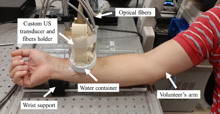
To understand the impact of melanin content on conventional amplitude-based PA images and investigate whether SLSC may reduce clutter, researchers — who collaborated with colleagues in Brazil who had previously used on of Bell’s algorithms — tested the forearms of 18 volunteers with skin tones ranging from light to dark. They used an imaging system consisting of a Nd:YAG laser (Brilliant B; Opotek) and a trifurcated optical fiber bundle (77,536; Newport) attached to a linear array transducer (L14-5/38; Ultrasonix) with a center frequency of 7.2 MHz and 128 elements.
The results showed that compared to the conventional PA images, using a signal-to-noise ratio — a scientific measure comparing signal with background noise — improved imaging for all skin tones when using SLSC while performing medical imaging. Of note, this technique was originally used for ultrasounds, but can be applied to PA imaging.

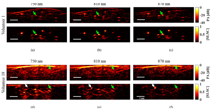
Referring to an image included in the study, researchers said, “The SLSC PA images in Fig. 4 contain reduced clutter and better visualization of the RA [radial artery] when compared to the amplitude-based PA images. In addition, the SLSC PA images qualitatively have good visualization of the RA and a low level of clutter artifacts, for both the light and darker skinned volunteers. This observation is further supported by the SLSC PA images in Figs. 4(d) to 4(f) improving visualization of a small blood vessel (white arrow) previously masked by clutter in conventional PA images.”
While the study wrote that skin tone bias did still exist to some extent in some of the SLSC PA images produced in the study, researchers focused on the bias reduction seen with SLSC compared to amplitude-based PA images, which they deemed as a success.
“The quantifiable bias introduced by skin tone variations was successfully mitigated with SLSC… with contributing factors that can be divided into optical considerations, spatial coherence influences, and clutter reduction benefits,” according to the study. “Regarding optical considerations, while both the concentration of melanin absorbers and optical wavelength affect skin PA signal amplitude, melanin concentration appears to be more influential than optical wavelength when considering the impact on skin PA signal coherence.”
With these new developments in PA technology, diagnosing, monitoring, and treating health issues in darker skinned individuals may now be more accurate, reducing previously established imaging inequity.
About the Author(s)
You May Also Like


.png?width=300&auto=webp&quality=80&disable=upscale)
