Bionic Vision Breakthrough Mimics Natural Eyesight
June 27, 2014
A step along the road to the restoration of full-fidelity sight was achieved by a team of Stanford researchers who have used the electrical stimulation of retinal cells to produce the same patterns of activity that occur when the retina sees a moving object.
|
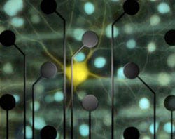
Chichilnisky and colleagues used an electrode array to record activity from retinal ganglion cells (yellow and blue) and feed it back to them, reproducing the cells' responses to visual stimulation. (Courtesy E.J. Chichilnisky, Stanford) |
The retina contains several cell layers. The first layer contains photoreceptor cells, which detect light and convert it into electrical signals. Retinitis pigmentosa and several other blinding diseases are caused by a loss of these cells. Many retinal prosthetics compensate for this loss by stimulating the retinal ganglion cell layer, the last layer of the retina before visual signals are sent to the brain. There are 1 to 1.5 million retinal ganglion cells inside the retina, of at least 20 varieties. Natural vision--including the ability to see details in shape, color, depth, and motion--requires activating the right cells at the right time.
The team focused their efforts on a type of retinal ganglion cell called parasol cells. These cells are known to be important for detecting movement, and its direction and speed, within a visual scene. When a moving object passes through visual space, the cells are activated in waves across the retina.
The new study shows that patterned electrical stimulation can do just that in isolated retinal tissue. "We've found that we can reproduce natural patterns of activity in the retina with exquisite precision," said E.J. Chichilnisky, PhD, a professor of neurosurgery at Stanford's School of Medicine and Hansen Experimental Physics Laboratory.
The study, "High-Fidelity Reproduction of Spatiotemporal Visual Signals for Retinal Prosthesis," has been published in Neuron. The lead author was Lauren Jepson, PhD, who was a postdoctoral fellow in Chichilnisky's former lab at the Salk Institute in La Jolla, CA. The pair collaborated with researchers at the University of California, San Diego; the Santa Cruz Institute for Particle Physics; and the AGH University of Science and Technology in Krakow, Poland.
There is a long way to go between these results and making a device that produces meaningful, patterned activity over a large region of the retina in a human patient," Chichilnisky said. "But if we can handle the many technical hurdles ahead, we may be able to speak to the nervous system in its own language, and precisely reproduce its normal function."
Refresh your medical device industry knowledge at MEDevice San Diego, September 10-11, 2014. |
The study was funded by the NIH's National Eye Institute, the National Science Foundation, the McKnight Foundation, the San Diego Foundation Blasker Award, and the Polish government.
Stephen Levy is a contributor to Qmed and MPMN.
About the Author(s)
You May Also Like


.png?width=300&auto=webp&quality=80&disable=upscale)