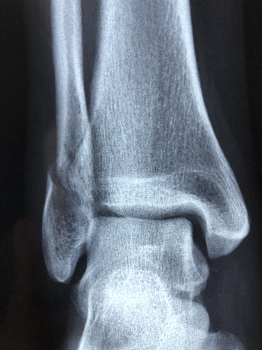
X-Rays Imaging Uses Color to ID Bone Fractures
Researchers at the University of Maryland, Baltimore County, and the University of Baltimore are testing this new method.
November 12, 2019

Researchers at the University of Maryland, Baltimore County (UMBC) and the University of Baltimore (UMB) are testing a new method of X-ray imaging that uses color to identify microfractures in bones. Microfractures were previously impossible to see using standard X-ray imaging. The findings associated with this advancement in color (spectral) CT imaging are published in Advanced Functional Materials.
Dipanjan Pan, professor of chemical, biochemical and environmental engineering UMBC, and professor of radiology at UMB, is the corresponding author of this new study. Looking ahead to the next generation of X-ray technology, researchers asked, "How can we detect a bone microcrack, something that is not visible using X-ray imaging?"
Researchers explained that to examine this question, they developed nanoparticles that navigate and attach specifically to areas where microcracks exist. Researchers call them "GPS particles." They started conducting this research at the University of Illinois Urbana-Champaign. The researchers have programmed the particles to latch onto the correct area of the microcrack. Once the particles attach to microcracks, they remain there, which is crucial to the imaging process.
The particles contain the element hafnium. A new X-ray-based technique developed by a New Zealand-based company MARS then take CT images of the body and the hafnium particles appear in color. This provides a very clear image of where the bone microcracks are located.
Hafnium is used because its composition makes it detectable to X-rays, generating a signal that can then be used to image the cracks. Researchers showed that hafnium is stable enough to be used in testing involving living creatures, and can be excreted safely from the body. The lab has not yet begun testing on humans, but the technology to do so may be available as soon as 2020.
As for other applications for spectral CT imaging with this hafnium breakthrough, the research suggests that this methodology could be used to detect much more serious problems. For example, in order to determine whether a person has a blockage in their heart, doctors often will perform a stress test to detect abnormalities, which comes with a significant amount of risk. One day in the near future, doctors may be able to use spectral CT to determine whether there is a blockage in organs.
About the Author(s)
You May Also Like


