Shattered Bones Get a Boost from Glass
February 1, 2007
R&D DIGEST
|
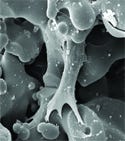
Within the glass scaffold, macropores and nanopores are interconnected. |
Glass that has interconnected pores and could assist in bone vascularization. The glass has potential as a biocompatible material for implants in orthopedic procedures.
According to Himanshu Jain, director of the International Materials Institute for New Functionalities in Glass at Lehigh University (Bethlehem, PA), the glass's porosity is the key to its use. He says it can be used to treat broken bones or osteoporosis. “The ideal treatment for diseased or damaged bone is to coax the body's natural bone tissue,” he says.
“Doctors do this by taking a bone graft from one part of a person's body and using it as a scaffold to stimulate bone tissue elsewhere to regrow.”
Likewise, he says, biocompatible glasses have been used as bone transplants. The glass is unique because it is porous at two scales. It contains nanopores that measure up to 20 nm in diameter, and macropores measuring 100 µm or wider. The nanopores facilitate cell adhesion and crystallization of bone's structural components, Jain says. The macropores allow bone cells to grow inside the glass and to form new blood vessels and tissue.
|
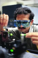
Director Himanshu Jain leads the international research project that has created dually-porous glass for use in orthopedic implants. |
The research team includes members from Lehigh, Princeton University, the University of Alexandria in Egypt, and the Instituto Superior Tecnico in Portugal. So far, the international team has been able to create glass that features both nanopores that have diameters of 5–20 nm and macropores.
To get there, the traditional melt-quench glass-making technique had to be refined. Members at the Alexandria facility developed a recipe for the powders that make up the glass. The materials are a mixture of silicon, calcium, phosphorus, and boron oxides. A chemical treatment then etches the glass to induce the desired porosity. They also used a sol-gel technique for glass-making that encourages the development of nano-sized pores. Then the team added a polymer to the solution.
The polymer caused a phase separation to occur parallel to the sol-to-gel transition, which helped overcome a significant obstacle, explains Jain. “Thermodynamically, the coexistence of nanopores and macropores is unstable in that the larger pores should absorb the smaller pores,” Jain says. “We have developed a material that defeats that expectation.”
Jain's Lehigh group is in the process of refining the fabrication process and investigating the mechanical properties and bioactivity of the materials.
In addition, another team at Lehigh is also measuring cells' interactions to the glass. The researchers have found that when the glass is attached to the damaged bone, a layer forms on the surface of the glass that has the same chemical composition as the natural bone.
The new material has been successfully tested in laboratory experiments, and the researchers are currently conducting in vivo tests at the University of Alexandria.
The researchers are also collaborating through the U.S.–Africa Materials Institute, which is headquartered at Princeton. The project is funded by the National Science Foundation.
Copyright ©2007 Medical Device & Diagnostic Industry
About the Author(s)
You May Also Like


.png?width=300&auto=webp&quality=80&disable=upscale)
