High-Resolution Sensor May Help Cancer Surgeons
July 18, 2006
Originally Published MPMN July 2006
EMERGING TECHNOLOGIES
High-Resolution Sensor May Help Cancer Surgeons
|
Side view of the device. |
|
Side view of a penny being pressed on the device. |
One of the trickiest decisions a cancer surgeon has to make is when to stop cutting. Stopping too soon may leave cancer cells in the patient’s body, and taking too many cells could also do unnecessary damage.
That decision could soon be made much easier, though, thanks to a high-resolution touch sensor developed by chemical engineers at the University of Nebraska–Lincoln. The sensor may allow surgeons to tell at the level of a single layer of cells whether or not they have excised a tumor in its entirety.
The engineers have developed a self-assembling nanoparticle device that has touch sensitivity comparable to that of the human finger.
“The touch resolution of the human finger is 40 µm,” said Ravi Saraf, the University’s Lowell E. and Betty Anderson professor of chemical engineering. “Using nanoparticles, we can attain resolution close to human touch, which is about 50 times better than what is out there today.”
Saraf explained that existing technology presents problems for use in minimally invasive surgery because the devices have low resolution, and are expensive and rigid, making them unsuitable for surgical applications.
He said the device that he and his doctoral student Vivek Maheshwari developed would be significantly cheaper because the device self-assembles at room temperature. It can also be made to cover an area of 1 square meter or larger, and is flexible enough to cover complex shapes.
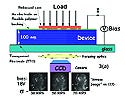
The device consists of alternating monolayers of gold nanoparticles 10 nm in diameter and cadmium sulfide nanoparticles 3 nm thick, separated by alternating layers of polymers that act as dielectric barriers. The manufacturing process is essentially a series of dip-coatings in various solutions with intermediate washing and drying processes. Saraf says the interactions between the materials at the atomic level is strong enough that they come together in a certain direction and a certain form, but weak enough that the nanoparticles can self-adjust an incorrect fit.
Gold and cadmium sulfide are conductive and semiconductive materials, Saraf says. “When you press on the device with an applied voltage across the thickness, that results in larger current and electroluminescent light from the semiconducting particle. By focusing the emitted light intensity from the cadmium sulfide particles or the change in local current throughout the device, you know how much pressure you have applied and how it changes over the contact area.”
As a demonstration experiment, Saraf and Maheshwari pressed a penny against a sample device and, using a charge-coupled device camera, they were able to decipher fine features such as wrinkles in Abraham Lincoln’s clothing.
“I am excited about this because I want to try to decipher cancer at the single-cell level,” Saraf says. “Because in some cases, cancer tissues are harder than normal tissues, if you take a tissue sample, put it on a glass slide and press on it, you would be able to see a cluster of just a few (cancer) cells with this method because it can sense down to about 10 µm. Surgeons will be able to know if they have taken out all of the cancer. If they haven’t, they’ll know where to make the next cut.”
Copyright ©2006 Medical Product Manufacturing News
You May Also Like


.png?width=300&auto=webp&quality=80&disable=upscale)
