Imaging Technique Could Unlock DNA Secrets
July 18, 2016
Harvard researchers have developed a new method for imaging molecules in DNA structures with high clarity and resolution.
Kristopher Sturgis
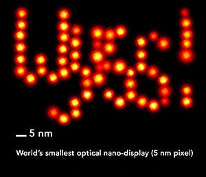
 The discovery, known as discrete molecular imaging (DMI), has enabled researchers at Harvard's Wyss Institute for Biologically Inspired Engineering to view individual molecular features that are distanced only 5 nm apart in a single molecular structure. Mingjie Dai, graduate student at Harvard and first author on the work, says that this discovery could provide valuable insights on the functioning of mechanisms at the molecular level.
"Many cellular and molecular processes rely on the collective working of many different molecules, in large macromolecular complexes or in complex molecular environment," he says. "DMI technology potentially allows scientists to investigate their composition and relative arrangement, and sheds light on their functioning mechanisms. The ability of visualizing and investigating these molecular machineries on a molecule-by-molecule basis provides doctors a way to investigate and diagnose diseases in the future."
This new technique adds to the group's DNA-PAINT platform--a collection of technologies that provided researchers with inexpensive, super-resolution microscopy that can take advantage of the transient binding of two complementary short DNA strands. The researchers attach on a strand to the molecular target that they aim to visualize, and the other strand is attached to a fluorescent dye. The group then repeats the cycle of binding and unbinding to create a defined blinking behavior of the dye at the target site, which can be exploited to achieve ultra-high resolution imaging.
The group was able to exploit the programmable nature of DNA molecule hybridisation, and use it to flexibly and precisely control the blinking temporal patterns on single biomolecular features.
The method opens up the possibility for researchers to study molecular conformations and heterogeneities in multi-component complexes, as well as providing a fast and easy method for the structural analysis of many samples.
"With this new power of resolution and the ability to focus on individual molecular features, DMI complements current structural biology methods like x-ray crystallography and cryo-electron microscopy," Dai says. "The higher power of resolution, and ability to focus on single molecular features provides a way to study single molecular features inside a big biomolecular complex, or in complex environments."
The discovery is just the latest in the advancement of microscopic methods, as researchers continue to extend the boundaries of image resolution at the molecular level. Yale University researchers recently reported the development of a new instrument that can enable scientists to image the tiny structures inside cells in three dimensional views. Much like Dai and his colleagues, the discovery out of Yale University demonstrates how techniques in microscopy continue to enhance the world around us at the molecular level, helping scientists achieve an even greater understanding of the structures and functions of cell biology.
As they move forward with their research, Dai and his colleagues intend to begin applying the technique to actual biological complexes, such as the protein complex that duplicates DNA in dividing cells -- a study that could one day lead to an enhanced understanding of the molecular signatures of disease, and could eventually lead to better diagnostics and treatment.
"Ultimately, our newly developed technology would provide researchers a way to visualize the discrete molecular components," he says. "This would help us understand their underlying role in versatile cellular functions, and provide doctors with a way to diagnose diseases based on molecular signatures."
Discrete Molecular Imaging from Wyss Institute on Vimeo.
Kristopher Sturgis is a contributor to Qmed.
Like what you're reading? Subscribe to our daily e-newsletter.
About the Author(s)
You May Also Like

.png?width=300&auto=webp&quality=80&disable=upscale)
