A Close-Up Look at Mauna Kea Technologies' Extreme Microscopy
October 1, 2012
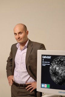
 Endomicroscopy pioneer Mauna Kea Technologies (Paris) has developed a tiny flexible microscope that can provide live, real-time images of internal human tissue. To learn more about the technology, we spoke with the company's president and CEO: Sacha Loiseau, PhD, who also co-founded the firm.
Loiseau, an astrophysicist who has worked at NASA, had a vision to take technologies used to probe the mysteries of the universe and use them to obtain microscopic images of the human body.
MPMN: Can you give us a summary of your technology and the applications of the Cellvizio technology?
Loiseau: We have been leading the charge for this new paradigm that is going to totally change the way physicians and surgeons perform procedures and treat patients. We've been focusing for many years on gastroenterology and have shown the power of optical biopsy for a growing number of indications. We are looking at a number of medical specialties to show that, as we were enjoying commercial success in gastroenterology, we could easily replicate the model to other medical disciplines. We want to explore put our probes into the hands of very skilled forward-looking surgeons to investigate its use to treat indications applications ranging from OB-GYN to ENT indications.
MPMN: How has your background in astrophysics prepared you to be the CEO of a medical device company? What parallels to you see between the two fields?
I would say the similarities [between those two fields] are myriad. The first is on the technology side. My specialty in astrophysics was designing and implementing new types of telescopes--optical technologies that enhance existing or future telescopes. The idea is that you are trying to look at very faint objects that are very distant to either get an image of it or measure it.
We think about what we are doing now in the human body: we are also trying to detect and image very faint objects--cellular structures that are extremely small and they are buried inside of tissue. We are often chasing objects in a very low-light and noisy environment and we want to make very beautiful images of the tissue. The technologies that we are combining to make this happen are really the same ones used in optical astronomy: we are looking at optics, electronics, mathematics, and mechanical and micromechanical issues [that are just as relevant for medicine as they are for astrophysics].
The technology we are using is based on the extreme miniaturization of advanced microscopic concept that has existed for decades. It enables very precise microscopic imaging using laser scanning of tissue instead of the traditional microscopy, which is based on using a lot of lenses. In our case, we don't magnify much. We use the laser to look at the tissue at the microscopic level and we make images based on what the laser is seeing on the tissue. We do this using optical technology that did not exist 20 years ago: fiber optics that are very thin pieces of glass where you can send light at one end and it exits at the other. We use at least 30,000 of them to scan an advanced laser through a small piece of tissue and take very precise images of the tissue. That's the basic concept.
There are other techniques we use that enable us to view the inside of tissue instead of just the surface. This is what has been invented on the bench top 50 years ago but never translated into a miniaturized objective.
Imagine that you have a closed book in front of you, and you can put an objective on the cover and it will show you page 100 without turning the page. That is what we do.
Another analogy that may be helpful here: If you take an old-fashioned flashlight and put it in the palm of the hand, if you turn it on, you can see through the other side glow red. Light goes through your entire hand. We use that concept to basically send light inside of the tissue. There is not a lot of light goes in and not a lot comes back. The trick is that we manage to detect very small amounts of light that can be detected at a precise location
We have taken this to the extreme because now we have these miniaturized objectives that are so small that we manage to fit them inside of a needle. We have a miniprobe that goes inside a 1-mm needle that is usually used to aspirate fluid. It can be used to, say, go into a pancreatic cyst or a lymph node or a solid mass. The surgeons aspirate the fluid that might contain cancerous cells. It is a bit of a game of finding a needle in a haystack. You are trying to find some cells that might be in this fluid that might be cancerous. It is not a good procedure in terms of yield. It is very hard to get something. People had been pushing us to put miniature objectives inside this needle to directly see what no other technology ever could, and that is what we've done with these probes that can go inside places where there has never been imaging technology. This really is extreme microscopy.
Brian Buntz is the editor-at-large at UBM Canon's medical group. Follow him on Twitter at @brian_buntz.
About the Author(s)
You May Also Like


