A New Treatment for Atrial Fibrillation?
Originally Published MDDI February 2006R&D DigestEngineers at Duke University (Durham, NC) hope to treat atrial fibrillation by combining ablation with three-dimensional ultrasound imaging in one catheter. Atrial fibrillation occurs when irregular cells trigger an abnormal heartbeat.
February 1, 2006
R&D Digest
Engineers at Duke University (Durham, NC) hope to treat atrial fibrillation by combining ablation with three-dimensional ultrasound imaging in one catheter. Atrial fibrillation occurs when irregular cells trigger an abnormal heartbeat.
“The first big advance is that we can actually do real-time 3-D ultrasound imaging at the tip of a catheter that's only 2 mm in diameter,” says Stephen Smith, PhD, professor at Duke's Pratt School of Engineering. “The second big advance is that we've added the capability of doing ultrasound ablation in the same catheter.” Such technology could be used for any application in which doctors are currently using radio-frequency ablation, according to Smith.
|
“Currently, ablation is done using radio frequencies, like microwave energy, from a separate catheter that goes into the heart,” Smith says. During the procedure, a doctor uses a fluoroscope to guide an electrode-tipped catheter to the heart. Radio waves are sent to the cells that produce the electrical signals that cause an irregular heartbeat. The method destroys these cells, but it can also overheat the tissue. In addition, fluoroscopy can't image soft tissue, making it hard for doctors to see the heart clearly.
The scientists' technique, ultrasound ablation, doesn't need to touch the tissue. Sound waves move through the blood and destroy irregular cells from 1 to 2 cm away.
|
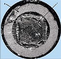
Figure 2. This model is about 4 mm in diameter. The imaging transducer (I) is encased in the catheter (A) so that real-time images may be taken. |
Researchers at the university have been making improvements to imaging techniques for a decade. Past work involved miniaturizing ultrasound probes containing an array of hundreds of transducers. When placed inside the esophagus, the probes can provide images of the heart.
Smith's group was able to fit nearly 200 cables into a 3-mm catheter. This enabled them to combine 3-D imaging and ablation in one catheter. “Right now there are no 3-D ultrasound catheters,” says Smith. “The only types of catheters available do 2-D ultrasound imaging.”
The technology could be used to isolate tissue so that a cardiac arrhythmia cannot spread to the rest of the heart. It would show real-time 3-D images of the heart at the source of the atrial fibrillation. For example, the device (Figure 1) might display two pulmonary veins in the left atrium. A doctor would be able to electrically isolate the pulmonary veins by putting a linear lesion around them.
The catheter is hooked up to a 3-D ultrasound scanner, which is about the size of a small refrigerator. The imaging transducers look like a checkerboard and are located inside the ring in the model shown (Figure 2). The checkerboard, or matrix array ultrasound transducers, creates a 3-D image. The ring simultaneously produces ablation, guided by the real-time 3-D image. “We're trying to produce real-time 3-D ultrasound imaging technology and ablation in the same catheter,” says Smith. “We've built three different prototypes.”
One model (see Figure 2) is about 4 mm, but the smallest one produced by the Duke team is 3 mm. This is small enough to use clinically, but just barely, says Smith. While further miniaturization of the device is a “worthwhile goal,” the team's target right now is to make the device produce better images and more-efficient therapy energy. The materials also need to be more durable and able to withstand commercial use. Such changes need to be adopted before animal studies are started, says Smith.—Maria Fontanazza
Copyright ©2006 Medical Device & Diagnostic Industry
You May Also Like


.png?width=300&auto=webp&quality=80&disable=upscale)
