January 25, 2013
Nanoparticles are among the most promising medical materials, with applications spanning imaging, diagnostics, drug delivery, and infection control. Nanoparticles, however, also have the potential to be toxic, altering cellular machinery, potentially altering gene expression and causing transgenerational damage. Their toxicity can also be used for good, however, to selectively target and destroy cancer cells.
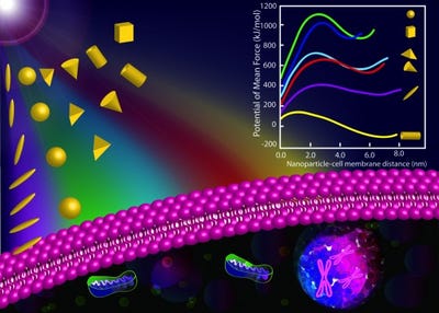
 Whether a given nanoparticle is toxic or not depends largely on a few principle physical variables: size, shape, and surface functionalization charge. In fact, those three variables were found in a recent study to influence the translocation of nanoparticles through cell membranes by as much as 60 orders of magnitude.
In the study, which was performed by researchers at Syracuse University, the translocation rates of cone-, cube-, rod-, rice-, pyramid-, and sphere-shaped nanoparticles through lipid membranes were analyzed. The most damaging nanoparticle configuration was positively charged, faceted, and rice-shaped. Particles with those attributes translocated instantaneously, causing substantial damage to membranes in the process. Negatively charged nanoparticles on the other hand are electrostatically repelled from the cell membrane.
The study, titled "Effects of nanoparticle charge and shape anisotropy on translocation through cell membranes," was published in Langmuir . It was authored by Shikha Nangia, Syracuse assistant professor of biomedical and chemical engineering and Radhakrishna Sureshkumar, department chair of the aforementioned department and physics professor.
Brian Buntz is the editor-in-chief of MPMN. Follow him on Twitter at @brian_buntz.
About the Author(s)
You May Also Like


