Modifying Metallic Implants with Magnetoelectropolishing
MACHINING
|
A finishing technique such as magnetoelectropolishing can complement machining operations. (Photo courtesy of iSTOCK PHOTO) |
Metals, their alloys, and intermetallic compounds are often used as medical device materials. Some metals are permanently covered with polymers that can deliver and controllably release drugs, others are coated with self-assembled hydrophilic or hydrophobic layers, and still others carry biological material (cardiovascular stents can carry endothelial cells, for example). But the majority of metals used in orthopedic and soft-tissue applications are for bare-metal devices.
An important niche of medical devices consists of cardiovascular and peripheral stents, which come in direct contact with blood. This creates a specific environment in which stents need to thrive. The most common problem with bare-metal stents is restenosis—when the body coats the stent with scar tissue and prematurely closes the treated artery or vessel. This problem led to the creation of drug-eluting stents, which seemed as though they would put bare-metal stents in the history books. Unfortunately, a new and more serious problem arrived: stent thrombosis. Several studies have linked this problem to drug-eluting stents, which, while fighting restenosis, simultaneously delayed and prevented proper endothelization of the stents. This delayed (and in most cases, never completed) endothelization is the source of clot formation.
One of the factors that determines a stent's bio- and hemocompatibility is its finish. The finish of stents and other bare-metal devices ranges from very coarse and porous to as smooth as possible. A common finishing process is electropolishing (EP), which delivers a very smooth finish and can be applied to delicate and intricate metallic devices. Medical device manufacturers have used this process for decades for orthopedic, dental, and soft-tissue implants and implantable devices. The EP process can be used as either an intermediate or a final step in production. This technique is often recognized for its brightening and leveling abilities, which include macro- and microsmoothing.1 An even more important property of the EP process is its ability to passivate and chemically modify a surface without changing the bulk properties of several commonly used materials in medical device production: 316L stainless steel, titanium and its alloys, and the intermetallic compound nitinol.
EP's passivation and modification properties have significant implications for medical implants, which come in direct contact with corrosive body fluids. What is not yet fully understood is which properties of electropolished surfaces are responsible for inhibition or reduction of initial bacterial adhesion. Some investigators point to minimized roughness, and others say modified surface chemistry, or a combination of both. Implant-associated infections are of great concern, because they can lead to serious complications such as soft-tissue damage, osteomyelitis, and implant rejection. Recent studies indicate that the electropolished titanium alloy Ti-6Al-7Nb may help minimize bacterial adhesion and lower the rate of infection.2
This article provides an update on research progress on a process called magnetoelectropolishing (MEP), which was introduced in March 2006.3 The process offers several advantages over other finishing techniques, including increased corrosion resistance, the ability to change the wettability of a surface, and improvement of a material's fatigue resistance.
Magnetoelectropolishing
The MEP process involves performing the standard EP technique under the influence of a constant magnetic field.4–6 In the EP process, an externally applied magnetic field works in two ways: the field either enhances or retards the dissolution rate of processed material. The dissolution rate depends on the strength of the applied magnetic field, but it is independent of the dissolved material's magnetic properties and the electrolyte used. Researchers have discovered that an externally applied magnetic field may either enhance or retard the dissolution rate during EP.3 This is due to the oxygen evolution regime, which influences the properties of the finished surface. Further research is required to understand why the magnetic field causes this change. In addition, a constantly applied magnetic field oriented parallel to the cathode surface during MEP suppresses hydrogen evolution. This minimizes the hydrogen embrittlement of magnetoelectropolished material.
MEP can be applied to any electropolishable materials. This article concentrates on experiments performed on two materials to compare the effects of MEP with other finishing methods. The materials were medical-grade 316L stainless steel, which is a prevalent implant material, and nitinol, which has also been used as an implant material. This article focuses primarily on nitinol, but touches briefly on others.
Stainless Steel
|
Figure 1. (click to enlarge) Polarization curves of 316L-stainless-steel samples in Ringer's solution after mechanical abrasive polishing (a), standard electropolishing (b), and magnetoelectropolishing (c). |
We investigated the corrosion resistance of 316L stainless steel in Ringer's solution, which is an artificially prepared human body fluid commonly used for corrosion resistance investigations. We used electrochemical techniques that involved potentiodynamic polarization curves and electrochemical impedance spectroscopy (also known as Nyquist plots). The polarization curves were measured after three finishing treatments: MEP, EP, and mechanical abrasive polishing (MP) using abrasive paper with grit size down to 1000. The corrosion rates were then computed based on these curves (see Figure 1). The resulting rates, which are presented in Figure 2, indicate that MEP offered the lowest corrosion rate, while MP resulted in the highest (EP was somewhere in between). Therefore, MEP may be the most advantageous process for modifying bare-metal implants.
|
Figure 2. (click to enlarge) Corrosion rate of 316L stainless steel in Ringer's solution after treatment with MEP, EP, and MP techniques. |
Nitinol
In recent years, interest in nitinol has grown steadily because of the material's remarkable combination of mechanical (pseudoplasticity and shape memory) and biocompatible properties. Nitinol owes its unusual mechanical properties to its ability to reversibly change between two crystalline structures. Its biocompatibility originates from its chemical composition. As an almost equiatomic binary intermetallic compound of nickel and titanium, nitinol undergoes spontaneous oxidation. The oxidation is key to its uses in orthopedic, dental, and biliary applications as well as to its hemocompatibility in peripheral stents and heart valves.7 The two main drawbacks of nitinol are the leaching of nickel ions, which can create allergic reactions, and suboptimal fatigue resistance, especially in devices that have to endure numerous bending and twisting cycles such as peripheral stents. As a result, the quest to develop a totally passive nitinol with improved fatigue resistance continues.
|
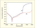
Figure 3. (click to enlarge) Polarization curves of electropolished (a) and magnetoelectropolished (b) nitinol in Ringer's solution. |
The EP of nitinol is performed in a number of alcohols and acid mixtures, as well as in salt electrolytes below and under oxygen evolution regimes. For this experiment, we used a proprietary electrolyte that is a mixture of nonhalogen acid and alcohol. A nonhalogen electrolyte was used instead of a halogen electrolyte because the nonhalogen electrolyte's composition rules out the possibility of incorporating Cl–, Br–, or F– ions into the oxide.8 These ions could cause passive film breakout and create sites of pitting corrosion. A constant external magnetic field below 1 T was applied to the EP system by neodymium ring magnets. The magnetic field was arranged parallel to the surfaces of the electropolished material. A standard parallel EP experiment was performed under the same conditions excluding the magnetic field in order to compare and evaluate differences between EP and MEP.
|
Figure 4. (click to enlarge) Comparison of nitinol's corrosion current after EP and MEP. |
Behavior in Ringer's Solution. High corrosion resistance is believed to be one of the main prerequisites for bio- and hemocompatibility. Manufacturers should create a protective TiO2 layer to minimize the Ni ions released to surrounding body tissue, which can sometimes trigger allergic reactions. An applied magnetic field selectively dissolves more nickel, and in doing so, it enriches the outermost layer of titanium.3 The corrosion currents, as proportional to the corrosion rates, were computed based on the polarization curves from this experiment (shown in Figure 3) and are presented in Figure 4. The curves indicate that MEP helps lower corrosion rates compared with EP. The decrease of corrosion current after MEP also lowers the Menne threshold of Ni ion release.9
|
Figure 5. (click to enlarge) Scanning electron microscope micrographs of electropolished (a) and magnetoelectropolished (b) nitinol. |
Scanning Electron Microscopy. Figure 5 shows micrographs of electropolished and magnetoelectropolished surfaces of nitinol. The electropolished surface shows a unique wavy pattern, which has also been documented by other researchers, that is not present on the magnetoelectropolished sample.10 The electropolished surface also shows more irregularities and imperfections.
Atomic Force Microscopy (AFM). The roughness measured by AFM for two scan sizes (see Figure 6) indicates that MEP has superior leveling properties compared with EP. The Lorenz force (the product of the magnetic field and current) could explain this to some extent. The mechanical effect of this force is the rotation of the electrolyte around the axis parallel to the direction of the magnetic field. The rotation of the electrolyte reduces the thickness of the diffusion layer (viscous layer).
|
Figure 6. (click to enlarge) Roughness of electropolished and magnetoelectropolished samples of nitinol, measured by AFM. |
Water Contact Angle and Surface Energy. Research suggests that the surface wettability of biomaterials on initial cell attachment and spreading is extremely important. Both mechanical (roughness) and chemical (functional groups) factors determine the wettability of a particular surface. It is broadly recognized that more-wettable (hydrophilic) metallic surfaces are more thromboresistant.11
|
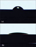
Figure 7. (click to enlarge) Image of water droplet deposited on electropolished (a) and magnetoelectropolished (b) nitinol surfaces. |
And in many cases, avoiding platelet thrombosis by using more-hydrophilic stents can minimize acute or subacute restenosis. Studies on metallic implants have concluded that for these types of implants, surface energy plays a more influential role on cellular adhesion and proliferation than surface roughness.12 Surface energy plays a role in the thromboresistance of hydrophilic materials; higher-surface-energy materials have stronger hydrating properties. The surface energy may also play a role in protein absorption (especially fibrinogen) and conformation, which lead to better thromboresistance by minimizing platelet adhesion.3 Compared with EP results, the static contact angle of nitinol after MEP (see Figure 7) shows a remarkable decrease, which correlates with the titanium enrichment of the outermost oxide layer.3 The wettability of this oxide is similar to that of oxide on pure titanium, which has long been recognized for its bio- and hemocompability. The change in wettability after undergoing MEP is not exclusive to nitinol—it also applies to other metallic biomaterials such as stainless steel and cobalt-chromium alloys.
Fatigue Resistance
One niche in which nitinol is finding more applications is in peripheral stents for superficial femoral artery (SFA), carotid, biliary, and urological applications. SFA stents are exposed to severe and continuous biomechanical forces such as flexion, torsion, compression, and elongation. These forces, in conjuction with the properties of nitinol stents themselves, often cause the stents to fracture.13 The problem could be the result of a poor surface finish, which is characterized by microcracks. Biomechanical forces cause the cracks to propagate and eventually fracture the stent. One of the surface-finishing techniques used to mitigate this potential problem is EP. A properly electropolished stent surface should be free of microcracks.
To see whether MEP could affect nitinol's fatigue resistance, a simple bending test was done in ambient atmosphere (according to Polish standard PN-75/M-80002, which defines specific conditions for performing bending tests). This study used two sets of nitinol wires with 0.5-mm diameters. Each set consisted of two wires cut to the same length. One set was electropolished, and the other was magnetoelectropolished, both with the same electrolyte under the same conditions. One cycle consisted of a 90Ëš bend in one direction, a return to the starting position, a 90Ëš bend in the opposite direction, and a return to the starting position. For both sets of wires, the magnetoelectropolished wires endured about 40% more cycles than standard electropolished wires before breaking.
|
Figure 8. (click to enlarge) Preliminary results of bending tests performed on nitinol samples after EP and MEP. |
Altogether, seven bending tests were performed on the electropolished and magnetoelectropolished samples. Due to discrepancies in the numbers of bendings after each test, the results of the tests with the two lowest and two highest numbers of cycles before breaking were abandoned. Thus, Figure 8 presents the results of three tests. Any microcracks that may have been present on the nitinol wires' surfaces were removed by the EP or MEP processes prior to the tests. It is well known that EP can introduce hydrogen to nitinol.14 It is possible that during MEP the evolution of hydrogen may be suppressed and a diamagnetic-species hydrogen may also be deflected from the nitinol. This would prevent or at least minimize hydrogen uptake, which would improve the fatigue resistance of the magnetoelectropolished sample.
The results of the corrosion and mechanical tests indicate that MEP is effective for treating metallic implants, and the process offers several advantages over standard EP. A magnetoelectropolished metal surface's improved properties may lie in the changed composition of the surface film formed after treatment.
|
Figure 9. (click to enlarge) Cr:Fe atomic ratios for 316L stainless steel in bulk, and after MP, EP, and MEP. |
The surface film of 316L stainless steel after EP consists of oxides and hydroxides of chromium and iron.15,16 The atomic ratio of Cr:Fe in the 316L-stainless-steel bulk is 1:3.65. After MP, the ratio is 1:2.10, and after standard EP, it equals 1:1.71. The 316L-stainless-steel surface layer after MEP revealed a ratio of 1:0.71 (see Figure 9). The surface film's higher content of Cr2O3 after MEP has a decisive influence on the material's corrosion behavior.
Conclusion
MEP can be applied to almost any electropolishable material, as was shown for medical-grade 316L stainless steel and nitinol. One of the most important properties of this technique is its ability to change the wettability of the process surface. It is worth noting that the wettability of metallic biomaterials can be adjusted by the strength of the applied magnetic field. This tailored, on-demand wettability could have many implications for bare-metal stents, which are used in various blood vessels that are exposed to varying shear conditions of flowing blood. With additional research, the manipulation of surface energy may be a strategy to use for directing the speed and quality of proper endothelization, which can help avoid or minimize restenosis. MEP can also improve a material's fatigue resistance, which could broaden nitinol's use in peripheral stents and other medical applications. Manufacturers can use this process to help improve production of their devices.
Ryszard Rokicki is the owner of ElectroBright (Macungie, PA), an electropolishing firm focusing on specialized metals. Rokicki can be contacted at [email protected]. Hryniewicz is the head of the surface electrochemistry division at the Koszalin University of Technology (KUT). Rokosz is an adjunct professor in the division of surface electrochemistry at KUT.
References
1. T Hryniewicz, “Concept of Microsmoothing in the Electropolishing Process,” Surface and Coatings Technology 64 (1994): 75–80.
2. LG Harris et al., “Staphylococcus aureus Adhesion to Standard Micro-Rough and Electropolished Implant Materials,” Journal of Materials Science: Materials in Medicine 18, no. 6 (2007): 1151–1156.
3. R Rokicki, “Magnetic Fields and Electropolished Metallic Implants,” Medical Device & Diagnostic Industry 28, no. 3 (2006): 116–123.
4. R Rokicki, “Apparatus and Method for Enhancing Electropolishing Utilizing Magnetic Fields,” U.S. Patent Application 2,006,124,472 (2006).
5. T Hryniewicz, R Rokicki, and R Rokosz, “Magnetoelectropolishing Process Improves Characteristics of Finished Metal Surfaces,” Metal Finishing 104, no. 12 (2006): 26–33.
6. T Hryniewicz, R Rokicki, and R Rokosz, “Metal Surface Modification by Magnetoelectropolishing,” in Proceedings of 16th International Conference on Metallurgy and Materials, Symposium D, Surface Engineering, D2 (Hradec nad Moravici, Czech Republic: Tangers, 2007), 1–8.
7. BG Pound, “Electrochemical Behavior of Nitinol in Simulated Bile Solution” (paper presented at International Conference on Shape Memory and Superelastic Technologies, Pacific Grove, CA, May 7–11, 2006).
8. MJ Graham, “Application of Surface Techniques in Understanding Corrosion Phenomena and Oxide Growth Mechanism,” Corrosion 59, no. 6 (2003): 475–488.
9. T Menne, “Prevention of Nickel Allergy by Regulation of Specific Exposures,” Annals of Clinical Laboratory Science 26 (1996): 133–138.
10. SA Summy, C Trépanier, and R Venugoplan, “Topographical and Compositional Homogeneity of Electropolished NiTi Alloy Surface,” in Proceedings of Society for Biomaterials 28th Annual Meeting Transactions (Mt. Laurel, NJ: Society for Biomaterials, 2002), 510.
11. IH Lee and HB Lee, “Platelet Adhesion onto Wettability Gradient Surfaces in the Absence and Presence of Plasma Proteins,” Journal of Biomedical Materials Research 41 (1998): 304–311.
12. NJ Hallab et al., “Evaluation of Metallic and Polymeric Biomaterial Surface Energy and Surface Roughness Characteristics for Directed Cell Adhesion,” Tissue Engineering 7, no. 1 (2001): 55–71.
13. DE Allie et al., “Nitinol Stent Fracture in the SFA,” Endovascular Today (July/August 2004): 1–8.
14. A Pelton et al., “Structural and Diffusional Effects of Hydrogen in TiNi” (paper presented at International Conference on Shape Memory and Superelastic Technologies, Monterey, CA, May 4–8, 2003).
15. T Hryniewicz, SJ Fernandes, and R Rokicki, “Comparative Auger Studies of 316L Stainless Steel,” PK+IST+EBM Project (Lisbon, Portugal, November 2006).
16. G Selvaduray and S Trigwell, “Effect of Surface Treatment on Surface Characteristics of 316L Stainless Steel” (paper presented at Materials and Processes for Medical Devices Conference, Boston, November 14–18, 2005).
Copyright ©2008 Medical Device & Diagnostic Industry
About the Author(s)
You May Also Like


.png?width=300&auto=webp&quality=80&disable=upscale)
.png?width=300&auto=webp&quality=80&disable=upscale)
