Studying Anti-Corrosion Surface Black Marking on Medical-Grade Metal with Ultrafast Lasers
How effective is laser marking for stainless steel, titanium, titanium alloy, and nitinol medical devices?
January 24, 2022
In medical device manufacturing (MDM), material and process are two key un-separable factors, particularly when it comes to biomaterials. According to a market analysis report from Grand View Research, “Biomaterials Market Size, Share & Trends Analysis Report By Product,” biomaterial market size is ~$106.5 billion in 2019 and expected continuously growth in the near future. Common biomaterials include polymers, metals, ceramics, and naturals. Given metal’s strength, toughness, fatigue resistance, flexibility, forgeability, and ductility, the material has become very important in fabricating various biomedical devices, especially for interventional devices.
Given metal’s strength, toughness, fatigue resistance, flexibility, forgeability, and ductility, the material has become very important in fabricating various biomedical devices, especially for interventional devices.
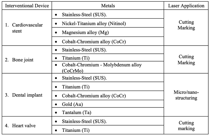
For such devices, material biocompatibility is essential for safety considerations. Thus, safe material processing is very critical to ensure that no change in material composition or properties occur in order to avoid safety and stability degradation. After decades of work, lasers have been accepted by the medical device industry and have become a standard for microfeature processing. For instance, bio-metal processing and device fabrication include laser (i) cutting, (ii) drilling, and (iii) surface modification. Table 1 shows major bio-metal based artificial human organ and applied laser applications. To comply with safety standards, ultrafast (ps or fs) lasers are ideal laser sources for device processing.
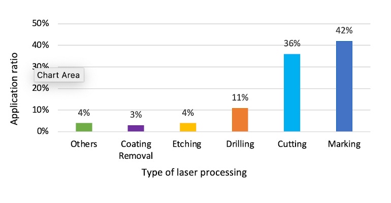
There is a huge demand for device marking. As seen in Figure 1 above, which shares statistics of various laser processing methods for >2000 samples processed in our application lab in the past 5 years, the data clearly shows that laser marking has been the most popular application.
The U.S. FDA has established the unique device identification (UDI) system to adequately identify medical devices sold in the United States from manufacturing through distribution to patient use.1 Besides its high visibility and contrast, medical device marking may have extra requirements due to its own particularity: markings must survive in the various harsh and even corrosive environments where medical devices may be used. In addition, markings should also survive after repeated autoclaving and/or sterilization processes for medical device re-use. All the above requirements put a great challenge on marking quality.
Laser marking is a quick and accurate process using the focused laser beam to generate labels or patterns on the material surface. It has unique advantages such as fast and permanent marking with high-resolution and non-contact marking, suiting lasers for metal back marking. 2-4 Especially, thanks to a pulse width shorter than that of metal electron-to-lattice coupling time (~1-2ps), non-thermal laser and matter interaction does not change chemical properties and makes an anti-corrosion black marking on medical metals possible.
According to studies on ultrafast laser-induced black metal marking process, two types of mechanisms have been discovered (as seen below in Table 2) depending on laser parameters: (i) Mechanism-1 is non-ablation surface modification/texturing, such as nano-ripples from laser-induced periodic surface structures (LIPSS) and black oxides (BO) from surface metal oxidation; (ii) Mechanism-2 is surface micro-ablation near the threshold to form micro/nanoparticles. This surface not only features a black surface, but it also offers hydrophobic properties.

In this article we share a systematic study on ps-IR laser-induced black marking on typical medical metals. As the experiment results show, marking on tested materials exhibits a good contrast and can survive in different corrosion environments as well as after the simulated autoclaving process. The experiment results suggest the laser is a promising marking technique well suited for the medical device industry and can be widely used in this field.
Experiment
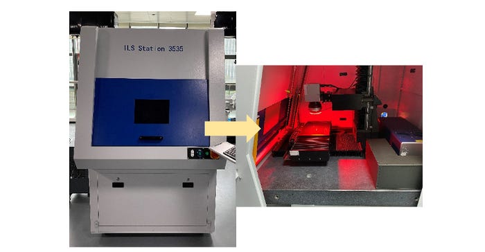
Four metals widely used in the medical field, stainless steel (SUS 316L), titanium (Ti), titanium alloy (T6Al4V), and nitinol (Ti-Ni), have been tested in this study using an all-in-one format AOPico IR (1064nm)/GR (532nm) and AOFemto IR (1030nm)/GR (515nm) lasers installed in AOC2000 series automated marking system (see Figure 2 and Table 3 below).

To test the anti-corrosion capability of the marking samples, the following corrosion tests have been carried out in sequence after the laser marking procedure:
Saline soap for 24 hours
7% CitriSurf 2050 at 50°C for 20 min
5% salt spray at 38°C (100 °F) for 72 hours
Besides the corrosion tests above, a simulated autoclaving test (home-used pressure cooker, 1hr/day, 7 days) was also carried out after the corrosion tests. The marking samples were examined in terms of the surface morphology and the material removal depth, through the optical microscope (Olympus BX51), the 3D laser microscope (Olympus OLS4100), and the SEM (Hitachi SU1510).
Result and Discussion
The marking samples after each procedure (marking, corrosion tests, and autoclaving test) have been examined, and it is found that there is no obvious difference before and/or after the corrosion/autoclaving tests in terms of the color, surface morphology, and the material removal depth, except where stated, and hence only the results after the corrosion/autoclaving tests are shown below.
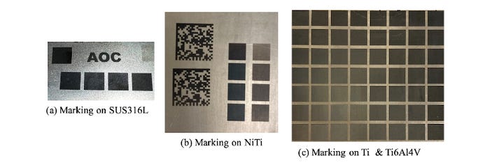
Shown in Figure 3a-c are some typical black marking samples on SUS316L, NiTi-alloy, and Ti and Ti6Al4V alloy plate, which passed corrosion and autoclaving tests. Simple test patterns and/or bar codes are used for marking with ps-IR and fs-GR laser, respectively.
The beauty of ps-IR laser-induced black marking is no obvious surface morphology change or material removal. A detailed surface analysis was investigated on the marked SUS316L and on Ti and Ti6Al4V sample.
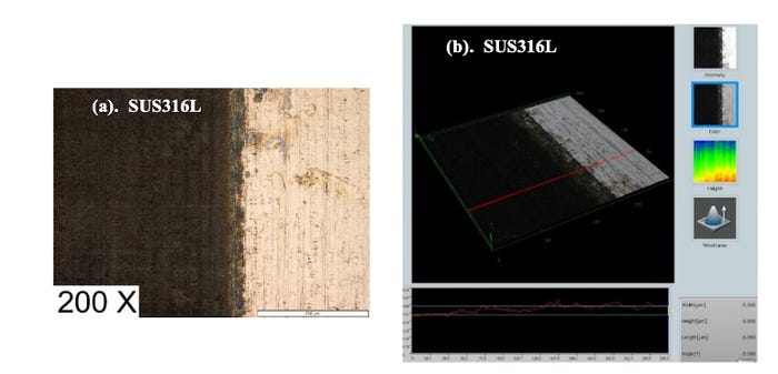
Figure 4a shows the surface morphology of a typical black marking area compared with the un-marking area, where no obvious surface morphology change can be observed, except the color. In addition, no laser beam moving tracks can be observed, suggesting it is a homogeneous marking procedure. Figure 4b shows the 3D laser microscope image: the material removal depth is ~0.85 um, which is the same as before the corrosion/autoclaving tests and indicates this is almost a damage-free process.
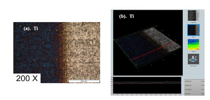
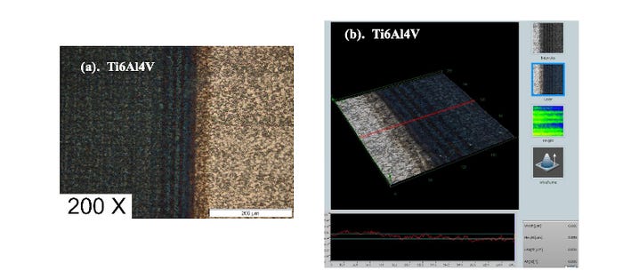
Similar results are also obtained on Ti and Ti6Al4V: the black marking survived after all corrosion/autoclaving tests. Figure 5a shows the typical black marking results on Ti compared with the unmarked area, where the homogeneous marking is also achieved with few changes on the surface morphology. Figure 5b shows the measurement of the surface ablation depth (~0.71 um) by 3D laser microscope, indicating a damage-free process. Figure 6a and Figure 6b shows the marking results on Ti6Al4V: the homogeneous marking is also realized on the sample surface with little material removal (removal depth ~ 0.86 um).
The underlying mechanism of the ps-IR laser black marking on the tested materials with the survival ability in the tested corrosion/autoclaving conditions deserves a detailed study. A possible explanation is that the marking process is a kind of pulse-heating-dominate treatment combining sub-µm ripple on the material surface with high peak power but low pulse energy, which is exactly the advantage of ultrafast lasers. Scanning electron microscope (SEM) is used to explore fine structures of laser treated zone.
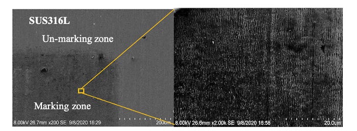
Figure 7 and 8 show SEM images of micro-structures of the marked SUS316L and Ti surface, respectively. It is clear to see sub-µm ripples on the marked SUS316L surface, but Ti surface. Thus, one reason could be due to a smoother surface of original SUS316L surface than Ti surface.

Laser marking is a gentle but continuous heat treatment that results in the permanent color change on the material surface with neither morphology change nor material removal. Such marking quality can be hardly achieved with a traditional long pulse laser, so the ultrafast laser is the right laser source for this special marking application.
Summary
In this paper, the ultrafast laser is used to generate a homogeneous damage-free marking on the tested materials, which can survive in different tested corrosion/autoclaving conditions. The test results suggest this anti-corrosion black marking technique is well suited for the medical device marking application and can be widely used in the medical field.
Reference
C. Leone, S. Genna, G. Caprino, I. De Iorio, "AISI 304 Stainless Steel Marking by a Q-Switched Diode Pumped Nd: YAG Laser," Journal of Materials Processing Technology. 210: 1297-1303, 2010.
A.J. Antonczak, B. Stepak, P. E. Koziol, K. M. Abramski, "The Influence of Process Parameters on the Laser-Induced Coloring of Titanium," Applied Physics A. 115: 1003-1013, 2014.
M. Gedvilas, B. Voisiat, G. Raciukaitis, "Grayscale Marking of Anodized Aluminum Plate by Using Picosecond Laser and Galvanometer Scanner," Journal of Laser Micro/Nanoengineering. 9(3): 267-270, 2014.
About the Author(s)
You May Also Like

.png?width=300&auto=webp&quality=80&disable=upscale)
