Implantable Chips and Advanced Materials Stimulate Research Efforts to Restore Sight
July 1, 1999
Medical Device & Diagnostic Industry Magazine
MDDI Article Index
An MD&DI July 1999 Column
Current efforts to develop a functional retinal implant are focused on returning useful vision to patients with retinitis pigmentosa and age-related macular degeneration.
Current research in bio-optics and bioelectronics could one day help restore a degree of vision to people afflicted by most common forms of blindness. Several engineering approaches are being explored to develop neuroprosthetic devices that can emulate normal vision to some degree. Much of this research involves the application of an innovative combination of technologies that have undergone remarkable progress in recent years, including advanced microprocessor systems, smaller and more-sensitive sensors, and new materials.
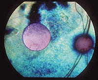 View of the fundus, showing the retina in which an artificial silicon retina has been implanted.
The retina is essentially tissue composed of several different cell types arranged in layers at the back of the eye. One layer contains the photoreceptors, the rods and cones, which take light signals that enter the front of the eye and convert them to neural signals. The most common retinal diseases, including retinitis pigmentosa (RP) and age-related macular degeneration (AMD), occur when the function of the photoreceptor cells is lost, becoming insensitive to light, while the rest of the cell types in the retina remain relatively intact. Because the function of photoreceptors is to convert light energy into neuroelectric energy, most of the current research is focused on artificially reproducing this activity to restore useful vision to the more than 10 million persons afflicted with RP or AMD worldwide.
"EYE OF NEWT" ADDS MAGIC TO PROTOTYPE ARTIFICIAL RETINA
In 1997, a group of researchers from the Institute of Physical and Chemical Research (RIKEN; Nagoya, Japan) and Nagoya University succeeded in developing an artificial retina design that combines semiconductor components with living nerve cells from newts. The novel device is based on use of a layer of cultured newt retinal cells positioned on a substrate of photoconductor elements. Light hitting the photoconductor substrate generates electrical signals that subsequently stimulate the nerve cells, the researchers indicate.
The researchers conducted computer simulations indicating that the photoconducters transmit signals to the retinal cells using a current of 0.1 to 1.0 mA. While the human retina relies on approximately 800,000 living cells that function as photodetectors, the prototype device uses only 64 silicon photodetectors. The research team noted, however, that the initial success of the prototype verified the potential of the device as a vision aid.
The group has built upon the information generated during its earlier success to develop a hybrid artificial retina that combines neural transplantation of human eye cells with a semiconductor device incorporating silicon photodetectors. The researchers recently noted that, using micromachining technology, a prototype microelectrode array was created for extracellular stimulation of nervous systems. The array consists of nine-channel bipolar electrodes, arranged in nine anode/cathode pairs in a 3 x 3 lattice. Each electrode has a 20 x 20-µm square shape. The distance between each pair is approximately 320 µm to reduce cross talk between pairs. In its next study, the group plans to evaluate the performance of the array during in vivo and in vitro extracellular stimulation experiments.
LASER POWERS IMPLANTED CHIP
In a separate project, a partial artificial retina is being developed by the Retinal Implant Project, a joint research effort being conducted by scientists from the Massachusetts Eye and Ear Infirmary (Boston), the Massachusetts Institute of Technology (MIT; Cambridge, MA), and Harvard Medical School (Boston). The researchers have designed and tested a prototype incorporating an "ultrathin microchip that can be surgically implanted on the retina," according to the MIT researchers.
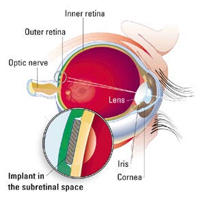 Surgically implanted under the retinal tissue, the artificial retina generates signals similar to those produced by the natural photoreceptor layer of the eye.
"The microchip serves to bypass the defective rods and cones by stimulating healthy ganglion cells directly with tiny electrical currents," the researchers explain. An 820-nm infrared external laser, along with a tiny camera, is mounted onto a pair of eyeglasses that capture visual images using the charge-coupled device (CCD). The images are converted to digital signals and transmitted by laser to the implanted chip. The chip then transmits the electrical impulses to the brain "by stimulating remaining healthy retinal ganglion cells using an array of electrodes surgically implanted on the retina," says John Wyatt, PhD, of the MIT department of electrical engineering and computer science. "The team has carried out electrical threshold measurements on nearly a hundred rabbit retinal ganglion cells to accurately determine the minimum amount of current and power needed for retinal stimulation. In addition, a number of short-term in vivo electrical stimulation experiments were performed, recording responses in the visual cortex to stimulation of small areas of the retina. Also, a large number of surgical experiments on laboratory animals were performed to develop improved techniques for implanting the device on delicate retinal tissue."
Although the laser has not been tested with the camera yet, the researchers recently completed a one-year experimental study of the biocompatibility of several implant materials, including silicone elastomer and hydrogels. "The next short-term objective is to refine the method for applying the silicone coating to the implant [because] even the smallest leak of salt from the eye into the implant would destroy function of the microchip," Wyatt explains.
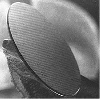 The ASR is powered solely by incident light and requires no external wires or batteries.
The goal of the research is to verify the brain's response to the implant, "first acutely, and then over progressively longer implantation periods," Wyatt says, adding, "This will involve repeated retest and redesign to reduce retinal trauma." Once this is accomplished, the researchers plan to "develop strategies to make the electrical stimulation as selective as possible for the desired cell types; design and build a second-generation implant capable of driving each of the stimulating electrodes separately; restore vision to animals that have been blinded by retinitis pigmentosa; and, finally, [start trials] with blind human volunteers."
BIOCOMPATIBILITY POSES AN ADDITIONAL CHALLENGE
Research led by Wentai Liu, PhD, a professor of electrical engineering at North Carolina State University (NCSU; Raleigh), and funded in part by a National Science Foundation grant is investigating the use of a prosthesis incorporating a laser-powered silicon bipolar contact artificial retina chip. The research is being conducted jointly with Mark S. Humayun, MD, PhD, and Eugene DeJuan, MD, of The Wilmer Ophthalmologic Institute at Johns Hopkins University (Baltimore).
"The major challenge is to have a miniaturized prosthetic device that is hermetically sealed, biocompatible, low power, and implantable," says Liu. "A complete prototype device is not finished, but major components have been done and tested." Liu also indicates that clinical testing of the prosthesis concept has been performed on 15 subjects to date—13 patients with end-stage RP and 2 suffering from AMD.
The prosthetic device comprises two subsystems. The first is outside of the eye to acquire, code, and transmit an image. The second, implanted within the eye, is designed to receive and decode image data, and then apply an appropriate stimulation pattern to the retina. The external portion of the system consists of a minicamera, video-capture circuit, and power and video-data transmission circuit mounted onto a special pair of glasses. Electrical power is transmitted wirelessly to the implanted chip using a two-wire coil configuration that functions similarly to a transformer.
The retinal chip within the eye has been designed to stimulate the retina in such a way as to form a 10 x 10-pixel image. According to the researchers, an improved version of this chip should be capable of sending operating-status data back to the glasses for monitoring. The chip specifications have been based on preliminary biological testing at Johns Hopkins University.
All of the components for the external subsystem have been designed and fabricated, and the final stage of assembly and testing is nearing completion. Says Liu, "Inside the eye, we are working toward a hermetic sealing package of the subsystem, which consists of retinal chip and retinal electrodes. Three generations of the retinal chips have been completely designed, fabricated, and successfully tested in function. Three kinds of electrodes, including platinum electrodes on silicone, platinum electrodes on polyimide, and platinum/iridium electrodes on silicon, are being designed and evaluated in vitro."
The researcher explains that, "Before the entire prosthetic device can be chronically implanted, all the components must be integrated in a biocompatible device. By the year 2000, we expect to have a complete prototype of the prosthetic device so that both long-term-based in vitro and in vivo tests in animals and volunteer human patients can be done."
The group's current research efforts are intended to overcome two challenges associated with the system—providing power to the chip, which is isolated within the ocular cavity, and fabricating the photoreceptors and electrodes on opposite sides of the chip to stimulate the retina. The researchers suggest that, because the cornea is transparent to laser light in any visible wavelength, such a laser could be used to power a chip equipped with photoconducting pixels. They further suggest that by fabricating the photoconducting photovoltaic cells, the photosensing or phototransistor array and the stimulating array could all be placed on a single side of the chip.
WORKING WITH AVAILABLE LIGHT
An artificial silicon retina (ASR) has been invented by brothers Alan Chow, MD, and Vincent Chow, an electrical engineer. The brothers are cofounders of Optobionics Corp. (Wheaton, IL), which is developing the technology. The silicon chip of the ASR is approximately 3-mm diam and 0.001-in. thick and functions as a subretinal microphotodiode array (SMA). The chip contains microphotodiodes that act as microscopic solar cells, each having its own stimulating electrode. The microphotodiodes are designed to convert the light energy from images to electrical impulses that stimulate the remaining functional retinal cells in patients with RP and AMD.
The researchers state that the novel design of the ASR allows it to be "powered solely by incident light, requiring no external wires or power sources." Surgically implanted under the retina, the ASR is designed to produce visual signals similar to those produced by the eye's natural photoreceptor layer. From their subretinal location, the ASR's photoelectric signals can induce artificial biological visual signals in the remaining functional retinal cells. These signals are processed and sent to the brain via the optic nerve. The researchers believe that the device is unique because it functions much like a solar cell, with no external connections and no power supply. The device is powered only by light that enters the eye.
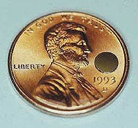 Optobionics' artificial silicon retina is approximately 3-mm diam and 0.001-in. thick.
Animal models implanted with the ASR during preclinical laboratory testing responded to light stimuli with retinal electrical signals and brain-wave signals. The induction of these biological signals by the ASR indicates that visual responses had occurred. Optobionics is presently working in collaboration with the Hines Veterans Administration Medical Center (Maywood, IL), the Louisiana State University Eye Center (New Orleans), and Stanford University (Palo Alto, CA) to enhance biocompatibility and function of the ASR so that human clinical testing may be initiated. To date, the chip will work in the eye for only a limited period of time, but development continues and tests in human eyes are anticipated within two years.
The researchers hope to define the nature of the interface between the retina and the subretinal microphotodiode array. "We've also noticed that the functionality of the device increases over a period of two or three months, peaks at a steady state for several months, and then begins to decline. So we need to find out what's behind that time course," he adds. Currently the device is capable of displaying only black-and-white images, they note, and works best in well-lit rooms. The researchers expect human testing to begin within two years.
"LEARNING RETINA" WOULD ALLOW FINE-TUNING OF IMPLANTS
Two interdisciplinary research teams founded in 1995 by the German Federal Minister for Research and Technology are focusing their efforts on techniques for electrically stimulating retinal cells to produce at least partial ambulatory vision in blind patients. The EPI-RET project, coordinated by Rolf Eckmiller, DrEng, at the University of Bonn department of computer science, developed a micromachined stimulator for implantation within the retinal tissue. The SUB-RET project, directed by Eberhard Zrenner, DrMed, of the University of Tübingen, has developed microphotodiode arrays for implantation underneath the retina.
Zrenner states that the SUB-RET group has succeeded in producing several prototypes of microphotodiode arrays that can be implanted into the subretinal space, and have tested the biocompatibility and stability of the prototypes. "We have developed two surgical techniques, one of which is very novel," says Zrenner. "We have implanted such chips in rabbits, rats, and pigs up to 14 months, and recorded visual potentials from the retina and visual cortex, elicited by infrared response." He adds that a number of challenges must still be resolved. "The chip works only with very bright light and presently we are developing a completely new generation of an active subretinal chip that receives external energy and uses the photodiodes just for switching this energy onto retinal cells." Zrenner notes that the group's progress to date shows promise for development of a functional device in the future. "We are convinced that the concept, in principle, is right. But of course we need a number of additional steps until we will arrive at a product suited to be implanted into a human being since this needs a number of additional developments regarding safety, efficiency, and accordance with regulations valid for medical devices."
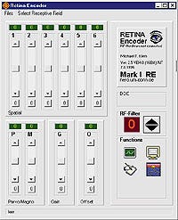 The EPI-RET project's Mark I retina encoder relies on computer simulations for RF-filter adjustment using neural nets and for monitoring the input and output signals.
Eckmiller, with Ralph Hünermann and Michael Becker, is conducting studies to develop a tunable retinal encoder (RE) for use with visual implants incorporating a retinal stimulator. This "learning retinal encoder," the EPI-RET researchers say, incorporates tunable spatiotemporal functions and a corresponding perception-based dialog procedure for the implant. Use of such a technique could allow the function of an implanted artificial retina to be optimized for restoring an individual patient's vision.
The initial version of the RE will be located outside the eye on the frame of a pair of glasses. Subsequent designs will allow the device to be embedded in a contact lens. The input of the RE comprises a photosensor array of approximately 100,000 smart pixels and 100 to 1000 technical ganglion-cell outputs that generate impulse sequences to elicit spike trains. An electromagnetic or optoelectronic wireless transmission channel is used to send the encoded ganglion-cell output to the implanted retinal stimulator, which is located adjacent to the retinal ganglion-cell layer.
The learning RE concept is based on using tunable receptive-field (RF) filters to stimulate the spatial and temporal receptive field properties of the retinal ganglion cells. Tuning the RF filters would entail generating RE modification signals to adjust specific parameters of the tunable filters so that the patient's perception of a visual stimulus most accurately represents the actual visual pattern. Although tuning the RF filters must be based on regained perceptions of blind subjects, the researchers recently proposed a method of pretraining the learning retina encoder using visual feedback provided by subjects with normal vision.
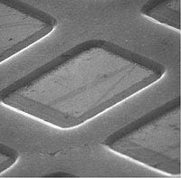 Individual microphotodiode located on an artificial silicon retina.
In response to the success of research efforts of the Bonn research team, Intelligent Implants GmbH (Bonn, Germany) was formed in the spring of 1998. The company intends to focus generally on development of the necessary technology platforms for learning neural prosthetic devices, among others. The company also intends to center its initial efforts on the commercial development of the learning retina implant.
CONCLUSION
Until recently, patients could be offered no hope of successful treatment to recover the sense of sight lost as a result of RP or AMD. The near-simultaneous development since the early 1990s of new materials with enhanced biocompatibility and of improved machining and circuit-design techniques is providing the tools that one day could allow vision to be restored to patients with these forms of retinal loss.
The majority of experts agree that the greatest hurdle to artificial retina development is inadequate funding. Neuroprosthesis procedures in the United States continue to be regarded as high risk, limiting the amount of available funds from both public and private sources. Eckmiller comments that, in addition to private funding, the ongoing research efforts at the University of Bonn have benefited substantially from government support as a leading project of the German neurotechnology effort.
Even the most advanced systems now in development, however, will have significant limitations at first. The tiniest of microphotodiodes remains quite large when compared to the natural cones of the human eye. Most researchers note that patients will achieve no more than blurred vision with present technology. And current chip designs are expected to allow only black-and-white vision because of limitations in wavelength detection. And there is some concern regarding potential damage from the implant to the remaining retinal cells. Once past the current threshold, and with adequate funding, continuing advances in materials and nanotechnology are expected to overcome initial limitations. Subsequent generations of implants are expected to provide restored vision with full-color images and with significantly greater resolution.
Gregg Nighswonger is executive editor of MD&DI.
Copyright ©1999 Medical Device & Diagnostic Industry
About the Author(s)
You May Also Like

.png?width=300&auto=webp&quality=80&disable=upscale)
