MX: Reducing Breast Cancer Bit by Bit
Breast cancer is the most common malignancy among women and the second leading cause of cancer deaths after lung cancer for women in the United States. Every year, 1.3 million women in the world are diagnosed with the disease, which takes the lives of 465,000 annually around the world, according to the American Cancer Society. Early detection by physical breast examination and mammography are the most important tools in the fight against the disease.
|
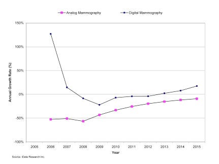
Growth rates, analog and digital mammography markets in the United States, 2005–2015 |
Mammography has been an invaluable method for detecting potentially cancerous abnormalities in the breast. While full-field digital mammography (FFDM) has diagnostic advantages over analog film mammography, both are limited by their two-dimensional projection capabilities.
A new technology substantially improves the diagnostic capabilities of standard digital mammography. Called digital tomosynthesis, this technology will be used in conjunction with FFDM in the coming years, substantially driving the global mammography market in the future. The benefits of digital tomosynthesis include lower patient recall rates, minimal retraining, reduced scan times, and small footprint.
Three Types of Mammography
Mammograms are images of the breast created from low radiation x-rays (1-3 milligrays, or mGy) that produce grayscale images. There are three types of mammography systems: analog, which uses film, computed radiography (CR), and FFDM. CR systems are film-based and retrofitted with a digital detector, but these systems are not considered fully digital. In addition, the overall radiation dose associated with FFDM at 1.86 mGy radiation per view is lower than that for conventional film, which averages 2.37 mGy radiation per view. Women with larger and denser breasts will experience the greatest reduction in radiation dosage. Younger women, for example, tend to have denser breast tissue compared with older women. FFDM detects over 25% more cancers than film mammography in perimenopausal women and in women under the age of 50.
Both analog and digital mammography produce images that are two-dimensional, and the tissues that absorb the x-rays are superimposed, potentially producing “summation shadows” and overlapping structures. The resulting mammograms can produce false negatives, because structures in the tissue may overlap and obscure the lesions in the image, making detection more difficult. Mammograms can also produce false positive results, where normal breast tissue mimics the appearance of abnormalities. In the United States, approximately 10% of screened women are called back for a second scan after an abnormality is detected. This takes an emotional toll on a patient because of the uncertainty regarding the diagnosis. In addition, secondary scans require considerable additional resources.
A team led by Daniel Kopans, M.D., pioneered the use of tomosynthesis techniques with GE Healthcare in the late 1990s at Massachusetts General Hospital (MGH) in Boston. The addition of digital breast tomosynthesis to FFDM is associated with a 30% reduction in the recall rate of patients who did not have cancer, according to a study of 125 patients at the University of Pittsburgh Medical Center. Digital breast tomosynthesis reconstructs 50 to 80 horizontal slices of breast tissue by taking successive images of the breast so that the tissue can be viewed and scrolled through. Eleven equidistant x-ray tubes sweep the compressed breast within a 50° angular range, acquiring two-dimensional scans and projecting them with a modified algorithm that produces three-dimensional images.
In addition, digital breast tomosynthesis requires very little retraining, as the procedure is similar to that of FFDM. The tomosynthesis system also occupies the same space as FFDM equipment, allowing hospitals to allot a similar footprint for a tomosynthesis upgrade as they do for their existing mammography equipment. Clinical benefits of digital tomosynthesis include the ability to see a number of reconstructed slices from different angles, increased visibility because of enhanced algorithms that concentrate on suspicious areas of tissue, and a reduction in scan time. Each examination takes seven seconds. In addition, a mammogram taken with digital tomosynthesis reduces patient discomfort because it requires less compression of the breast.
The Rise of Digital Tomosynthesis
The first digital mammography systems with breast tomosynthesis capabilities were installed in the Netherlands in 2008. Access to digital mammography continues to increase as facilities upgrade their systems from film to digital. As of 2009, the number of clinics equipped with digital tomosynthesis was very small. However, the improved diagnostic capabilities and cost reductions associated with lower false positive rates and patient recalls make digital tomosynthesis a worthwhile investment for healthcare facilities and an important technology for medical device manufacturers.
In Japan, many healthcare facilities are converting from film-based mammography systems and computed radiography systems to full-field digital systems. The conversion from film to digital technology was slow at first because digital photos did not previously offer the same resolution as film. However, digital mammography has now surpassed film in its resolution, convenience, and visualization capabilities. Digital mammograms do not require film developing because x-ray images are collected on a digital detector. Digital images can also be archived, easily sent to other healthcare facilities, and viewed as 3-D scans of breast tissue with the aid of tomosynthesis.
The market for digital mammography capital equipment declined in 2008 and 2009 as a result of the global economic recession. In addition, hospital budgets were constrained in 2009 as governments reined in healthcare spending. As a result of this economic uncertainty, purchases of premium one-time investments, such as capital equipment, were delayed during this period.
While the global digital mammography market was negatively affected by the recession, unit growth is expected to recover by 2015 and average selling prices are expected to rise as technological advancements such as digital tomosynthesis become more common. By 2015, the global market for digital mammography is expected to return to prerecession levels, exceeding a value of $679 million.
Boomers To Drive Digital Growth
The aging population of women, particularly baby boomers entering their 60s, and the increased use of digital breast tomosynthesis will drive the global growth of digital mammography. Digital breast tomosynthesis systems have been available in Europe and Canada since 2008. As of June 2010, FDA had yet to approve the systems for U.S. use, but they are available for research purposes. In late September 2010 FDA is scheduled to review breast tomosynthesis applications of Hologic’s Selenia Dimensions system, bringing the technology closer to premarket approval in the United States.
Overall, the clinical benefits to patients and cost savings to hospitals will encourage the acceptance of digital tomosynthesis. As a result, manufacturers of digital mammography equipment are expected to sell tomosynthesis as an upgrade to facilities with digital mammography systems, and digital breast tomosynthesis may ultimately replace mammography in the future. These trends will lead to double-digit growth rates in the U.S. and European breast imaging markets by 2015.
Kamran Zamanian is the head of research for iData Research (Vancouver, B.C.), an international market research and consulting group specializing in the medical device, dental, and pharmaceutical industries. Imelda Nurwisah is a research analyst for the firm. This article is excerpted from the study, Markets for Women’s Health Devices 2009. Additional information is available at 866-946-3282 or [email protected].
About the Author(s)
You May Also Like


.png?width=300&auto=webp&quality=80&disable=upscale)
