How Tiny Mirrors Could Boost Imaging of Living Cells
July 20, 2016
Researchers attempt to grow cells on tiny mirrors and image them with super-resolution microscopy, providing a three dimensional view of cells at the micron-scale for the first time.
Kristopher Sturgis
|
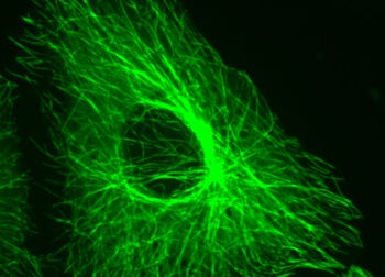
This Vero cell grew on the surface of a mirror, after which researchers fluorescently stained it to show the microtubules in the cell's cytoskeleton. (Image courtesy of Georgia Tech) |
U.S., Chinese, Austrialian researchers created the new technique, which is meant to address a longstanding problem in the community of cell biology: viewing cells in three dimensions with clear resolution in each dimension.
This improved view of living cells could help researchers differentiate between cell structures that appear close together, but actually exist relatively far apart within the cell. This kind of information could provide new details on cell behavior, and how diseases arise within them.
Traditionally, cells have been grown on transparent glass slides for microscopy observation, but this new technique aims to take advantage of the properties of light to create interference patterns as they pass through a cell toward a tiny mirror, and then back through the cell after being reflected. The interference patterns provide enhanced resolution in the Z-axis, which is what scientists see when they look directly into a cell perpendicular to the slide.
"Our idea came from a simple hypothesis," says Peng XI, professor in biomedical engineering at Peking University in China and one of the authors on the work. "Usually in super-resolution microscopy we need high depletion power to generate better resolution. However, the light only passes once through the specimen, and is then wasted by forward propagation. We thought that if we place a mirror behind the specimen, then the forward propagating laser can be reflected back, which is equivalent to increasing the power twice."
Xi says that previous efforts provided a blurred resolution akin to reading a newspaper printed on transparent plastic. To offset this, they decided to place a mirror beneath the specimen to generate a focal point, which essentially brings everything into focus with crystal clear resolution. Xi says that with this simple tool, biologists can begin tackling many of the interesting phenomena surrounding cell behavior that were invisible in the past due to poor resolution.
"This work brings both lateral resolution enhancement and axial narrowing to STED microscopy, therefore many fine subcellular structures, which cannot be resolved by conventional microscopy due to the fact that biological cells cannot tolerate high laser power, can now be visualized," Xi says. "This means that we won't be restricted to the superficial cell membrane, but rather enables us to penetrate up to 100 nm into the cell."
Xi and his colleagues hope that their new tool can help modify existing microscopy methods to provide clearer images of living cells.
"I think this method will be widely used in biological cell studies," Xi says. "Luckily, the study of subcellular structure is an important area in biology, and I believe our technique will play an important role in that. The application of this means is very straightforward. It is fully compatible with current commercial confocal and STED techniques, and we are looking forward to potential collaborations toward the commercialization of this simple technique."
Kristopher Sturgis is a contributor to Qmed.
Like what you're reading? Subscribe to our daily e-newsletter.
About the Author(s)
You May Also Like


.png?width=300&auto=webp&quality=80&disable=upscale)
