Medical “Lightsabers” Take Laser Precision to the Next Level
Theodore H. Maiman invented first working laser in 1960 while at Hughes Research Laboratories in Malibu, California. Soon after that, Maiman’s assistant Irnee D’Haenens quipped that the laser was “a solution looking for a problem.”It goes without saying that, over the past five decades, many applications or “problems” have been identified; lasers are used in countless applications—many of them in medicine, where they are used in everything from laser eye surgery to cosmetic treatments.
May 30, 2012

The use of medical lasers could be further expanded as their precision is improved. At present, lasers’ risk of damaging healthy tissue limits their application when used to target sensitive tissues such as those found in the brain, throat, and digestive tract.
To address this issue, a team of scientists at the University of Texas at Austin (UT Austin) has developed a flexible endoscopic device equipped with a femtosecond laser scalpel that can remove target tissue while sparing healthy surrounding cells. The technology could lead to a paradigm shift in the clinical use of lasers, says Adela Ben-Yakar, who heads the research at UT Austin. “It will enable new treatments as well as [improve] the current ones.”
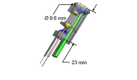
The medical "light saber" emits bursts lasting only 200 quadrillionths of a second and is equipped with a mini-microscope that enables high precision for delicate procedures. The current endoscope probe package measures 23 millimeters in length with a circumference of 9.6 millimeters, enabling it to fit into large endoscopes.
The packaged endoscope overlaid with the optical system. Image courtesy of Ben-Yakar Group, University of Texas at Austin. |
Ben-Yakar got involved in this research nearly a decade ago while doing postdoctoral research at Stanford University. “I was studying the laser interaction of materials and they were very precise and very clean. They could make very precise and clean cuts,” she says. “And then I started using them in cells and in biological tissues. I saw that the biggest impact really would come in biology and medicine,” she continues. “I got excited about this project and [the prospects of] bringing it to the clinic eventually.”
The initial applications that the researchers are targeting include eye surgery, repairing the vocal cords, and the removal of small tumors in the spinal cord or other tissues.
|
An image taken with the probe's two-photon fluorescence microscope shows cells in a 70-micrometer thick piece of vocal cord from a pig. Image courtesy of Ben-Yakar Group, University of Texas at Austin. |
Ben-Yakar estimates that the device will be ready for commercialization in about five years. “We will need a couple of years of animal studies and also a couple of years of clinical studies beforehand,” she says.
One factor that may help facilitate regulatory approval is the fact that femtosecond lasers are already in use clinically. “Of course, for specific applications, we will still need to get FDA approval but that may be a Fast Track approval since the LASIK femtosecond has already been approved for eye surgery,” she says. “That hopefully should pave the way for it to be used for different applications.”
A Laser that Can ‘See’ as well as Cut
The technology could potentially be used for imaging applications as well. “It can be used to ‘see’ what is happening within the target tissue with high resolution,” Ben-Yakar says. “The doctor would have information about what is happening inside of the tissue while cutting the surface of the tissue.” This could prove to be especially useful for application such as spinal cord surgery, where a doctor must be very careful in removing unwanted tissue because of the risk of severing the nerve cord.
Ben-Yakar says that when she explains this functionality to surgeons their eyes light up and they say: “That is what we would like to have. A tool with a single laser that can see as well as cut. It may, however, be longer than five years before this imaging functionality is available commercially.
Brian Buntz is the editor-at-large at UBM Canon's medical group. Follow him on Twitter at @brian_buntz.
About the Author(s)
You May Also Like


.png?width=300&auto=webp&quality=80&disable=upscale)
