Laser Microfabrication Takes on Diagnostic Consumables: Part 1
October 19, 2015
Advances in laser technology is leading to a wave of progress in diagnostic instruments.
Glenn Ogura, Resonetics
The trend to miniaturize diagnostic instruments leads to many benefits for patients and caregivers. It opens up opportunities for treating patients closer to the home. It simplifies testing by quickly putting the raw sample into the instrument and getting the result almost immediately, avoiding extra time to prepare the samples prior to testing. Miniaturized instruments also enable multiplexing tests together, which not only saves time, but also improves patient care and is more cost-effective than conventional methods. Miniaturized diagnostic platforms such as biochips and microfluidic chips are able to handle smaller volumes of samples and reagents for sample preparation, cell capture, and immobilization and testing.
To drive down costs and improve performance, many diagnostic instruments are now being made from glass and polymers, rather than silicon or ceramic. This switch creates new applications and opens the door to new fabrication methods. Laser micromachining is seen as one of those enabling microfabrication methods for the machining of consumables in fields such as molecular diagnostics, biosensors, single cell analysis, sequencing, and point-of-care.
Part 1 of this article discusses how the specialized capabilities of laser micromachining can be used for fabricating diagnostic consumables. Part 2 will provide an overview of the fundamentals of laser micromachining, and also compare laser micromachining with traditional methods.
Diagnostics Applications Requiring Laser Micromachining
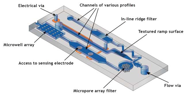
Years ago, biochips and microfluidic chips were fabricated from silicon, but today these chips are more often made from glass and polymers. In microfluidic chips, very small volumes of fluid (microliters to femtoliters) are transported, mixed, separated, and processed. See Figure 1. In biochips, device features include sample preparation, analyte collection and detection and data reporting.
|
Figure 1: A diagram of a multi-functional microfluidic chip. Note: This sketch is not intended to represent a real device; it is or illustration purposes only. |
Micropore Array Filter
The preparation of samples for a diagnostic test can be complex and time-consuming; it depends on the type of sample, such as whole blood, buccal swab (inside wall of cheek), urine, tissue, bacterial cultures, serum, stool, etc. The use of micropore array filters for sample preparation purifies the sample and improves the sensitivity of the test. These filters are an array of tiny laser-drilled holes (as small as 1 micron in diameter) in a polymer such as polyimide. Applications include diagnostic assays for flow control of reagents, blood filtration and blood separation for measurement of red blood cell (RBC) deformability, leukocyte removal, rare cell detection, and DNA sequencing.
As an example, for whole blood testing, the red blood cells must be separated from leukocytes (white blood cells) and other cells. The circulating tumor cell (CTC) assay is an emerging application involving the detection, analysis, and enumeration of rare cancer cells. Next generation CTC companies are focusing on how to capture the cells and then determine the potential for these cells to metastasize. These micropore array filters are ideal for whole blood filtration and rare cell capture as the tiny holes only allow small cell fragments to pass through.
Laser micromachining is useful for microfluidic chips because its parallel processing allows thousands of capture locations to be fabricated onto a chip, which then speeds up the analysis.
|
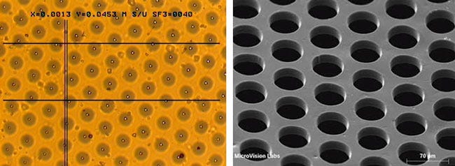
Figure 2A. A micropore array in polyimide is shown on the left. Figure 2B. On the right, a micropore array in polyimide. (1 micron diameter holes) (50 micron diameter holes) |
Figure 2A illustrates a laser-drilled array of 1-micron diameter holes in a polyimide film, used as a sample preparation filter for such applications as the creation of emulsion for a polymerase chain reaction (PCR) instrument. The consistent hole size, shape, and distance between holes create a more predictable PCR emulsion to improve the efficiency of molecular diagnostics and sequencing.
Microwell Array
For standard diagnostic tests, the actual reaction with the sample takes places inside a bioreactor.
As mentioned, there are market drivers to multiplex tests to reduce the diagnosis time and for the tests to be conducted outside the centralized hospital laboratories. As a result, miniature bioreactors are being developed that consist of arrays of blind micro wells in low fluorescence polymers, allowing hundreds of "mini tests" be simultaneously conducted.
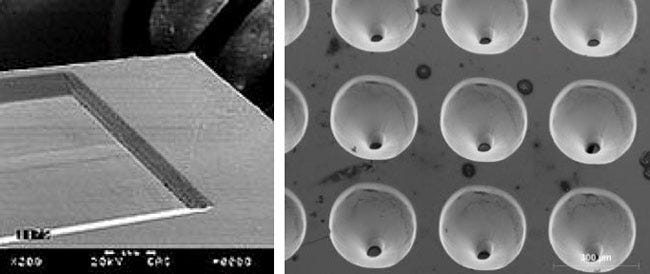
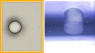
Lasers provide excellent depth control when fabricating microwells, as every laser pulse can remove material at a depth rate on the order of 0.1 micron per pulse. By controlling the number of laser pulses, the depth of each microwell in the array is controlled and the bottom surface of the wells is preserved (Figure 3A).
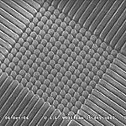
Lasers can also create custom hole profiles (e.g., a trumpet shape) in a microwell with great fidelity so that the analyte can more easily fill the bioreactor (Figure 3B). Lasers can also texture the surface of the bioreactor (Figure 4) to change its wettability properties.
|
Figure 3A. Blind well in polyimide (shown on the left). Figure 3B. Microwell (75 microns diameter) in COP film, |
Access to sensing electrodes
|
Figure 4. Micro-textured ZnSe. Courtesy of Optec s.a. and CSL. |
For diagnostic applications such as biosensors for the detection of glucose, DNA, antibody, immunoassays, or other cell-based sensors, the portal window in a cannula (Figure 5) or chip is a key design feature. It's critical that there is a consistent measure of analyte reaching an electrochemical or optical sensor embedded inside the cannula; the area of the portal window often is controlling the sensor signal level.
Flow Vias
Flow vias serve as an entry point for the analyte, such as liquid biopsy, into a microfluidic chip. Etching, CO2 lasers, and mechanical drilling are the most common methods to fabricate holes into the glass cover. As microfluidic chips become smaller, however, the individual layers become so thin (100 microns or less) that traditional methods do not work. Mechanical drilling leads to cracking and excessive debris, intolerable for cell transport systems. Powder blasting is limited to glass of typically 500 microns or thicker. HF etching may be prohibitive due the incompatibility of the chemicals with the patterned circuits on the chip or the isotropic nature of liquid etching.
|
Figure 5. Oval port window in nylon (4 mm in length) |
Laser micromachining overcomes the challenges and drills flow via holes in a single step in glass or polymers as small as 1 micron in diameter (see Figures 6A, 6B) with minimal debris or recast on the top surface.
The emergence of laser singulation of glass without material ablation opens up opportunities for ultrafast lasers to drill via holes, etch patterns and dice into individual glass chips. Applications include diagnostic platforms for sequencing, drug discovery and molecular diagnostics.
|
Figure 6A. Glass via, 200 microns diameter (shown on the left. Figure 6B 700 micron diameter via in 1 mm thick glass on the right. |
Electrical Vias
Electrical vias provide electrical conduit from one layer to another. For example, a via may connect the electrical signal from a diagnostic chip to a metal contact pad for external testing or monitoring. The metal pad can also serve as a landing pad for the target analyte.
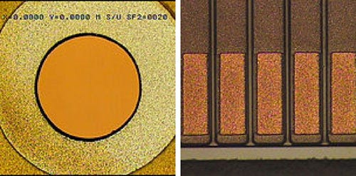
For many years, lasers have been a workhorse for the drilling of microvias (See Figure 7A) in electronics such as mobile phones. However, laser micromachining can also expose contact pads (Figure 7B) by selectively etching away the polymer coating without damaging the metal surface. This is because, for a properly selected laser, the damage threshold of metal is much higher than that of polymers; by over-pulsing, the laser process guarantees removal of 100% of the polymer coating from the surface of the metal pad. This direct process is more efficient than the combination of masking, deposition and cleaning, and offers the advantage of removing thicker coatings not possible with plasma cleaning.
|
Figure 7A. Laser-drilled via Figure 7B. Exposed gold electrodes. |
DNA microarrays (or DNA chips) operate when DNA probes are placed on the sensor area. Compared to photo lithography or mask deposition, laser micromachining is a single-step direct process that can selectively ablate polymer coatings from electrodes to permit attachment of the DNA probes that are constructed to be complimentary to the target nucleotides. Laser micromachining provides the benefit of saving multiple manufacturing steps, which in turn drives down costs.
Microchannels
A microfluidic chip is a tiny, integrated lab-on-a-chip consisting of a liquid handling system and a biochemical-processing device. Very small volumes of fluid (micro liters to femto liters) are transported, mixed, separated, and processed in a network of micro-channels.
Micro-channels can be fabricated in two ways. The lamination method involves sandwiching of a usually thin patterned middle sheet (die cut, ballistic punched or CO2 laser-cut) between two thicker plates to produce the desired channel structure. Cuvette designs can be a relatively simple three-layer construction or consist of more layers. When channel widths decrease to the 100-125 micron ranges, then die cut or ballistic punch are not feasible. In that case, the individual device layers are fed through a web-based converter composed of a CO2 laser directed by galvanometers onto the fast-moving web. The laser cuts out the pattern directly, offering the benefit of smaller channel dimensions and accelerating the development cycle.
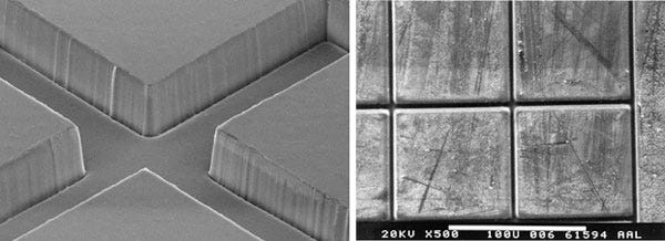
Alternatively, blind micro-channels can be etched directly into the chip using photolithography or wet or dry etching, which are ideal for silicon or glass platforms. For polymer-based chips, however, laser micromachining is an alternative to molding for certain applications, especially if the chip design includes through holes less than 150 microns in diameter. Lasers can create smooth-surfaced blind channels because the laser's etch rate is very low. For some microfluidic designs, microchannel dimensions are below 10 microns, which perfectly suits laser micromachining. For channel intersections, a mask projection method is preferred (see Figure 8A) so that the entire feature is machined at one time, avoiding a double-exposed intersection caused by the laser writing in both directions.
|
Figure 8A. Polyimide channel (50 x 50 microns) Figure 8B. Borosilicate channel (5 x 5 micron) |
In-line ridge filters
|
Figure 9. Laser-etched grooves |
For cell analysis instruments such as flow cytometry and cell counting, individual cells are placed in a single-file line so that they can be individually interrogated. See Figure 9. To prevent large particles from clogging a critical flow orifice, the in-line ridge filters use parallel channels instead of holes. Laser can etch these blind channel formations cost-effectively by machining multiple channels at one time.
Textured ramp surface
The wettability properties of diagnostic chips determines how they transport fluids to improve the efficiency of a miniature bio-reaction. Although surface modification can be accomplished by bead blasting, plasma or other methods that randomly modify the surface, laser micromachining offers the benefit of being a non-contact process that can machine repeatable micro-patterns at well-controlled locations. See Figure 10.
|
Fig 10. Surface modification of polymer |
Diagnostics is a critical field in the life science industry as instrument and reagent companies strive to develop assays to identify and treat illness in the early stages. Their end goal is assays that make all diseases totally preventable. As diagnostic instruments grow increasingly smaller, new applications emerge requiring microfabrication techniques. Laser micromachining is seen as an enabling technology for the making of diagnostics consumables in the fields of molecular diagnostics, biosensors, single cell analysis, sequencing and point-of-care.
This article will be continued in a Part 2, which will compare laser micromachining with traditional methods, and also provide an overview of the fundamentals of laser micromachining.
Glenn Ogura is the SVP Market Development at Resonetics (Nashua, NH).
Stop by Resonetics booth (#634) at BIOMEDevice San Jose, December 2-3. |
About the Author(s)
You May Also Like

.png?width=300&auto=webp&quality=80&disable=upscale)
