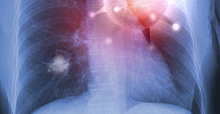
Chest X-rays Could be Key in COVID-19 Detection
The findings from the LSU radiologists were published in Radiology: Cardiothoracic Imaging.
September 3, 2020

A team of Louisiana State University radiologists investigated the usefulness of chest x-rays in COVID-19. Researchers found these X-rays could aid in a rapid diagnosis of the disease, especially in areas with limited testing capacity or delayed test results.
The research was published in Radiology: Cardiothoracic Imaging.
The radiologists conducted a retrospective study of nearly 400 persons under investigation (PUI) for COVID-19 in New Orleans. They reviewed the patients' chest X-rays along with concurrent reverse-transcription polymerase chain reaction (RT-PCR) virus tests. Using well-documented COVID-19 imaging patterns, two experienced radiologists categorized each chest x-ray as characteristic, nonspecific, or negative in appearance for COVID-19. The radiologists found a characteristic chest x-ray appearance is highly specific 96.6% and has a high positive predictive value of 83.8% for SARS-CoV-2 infection in the setting of a pandemic.
"The presence of patchy and/or confluent, band-like ground-glass opacity or consolidation in a peripheral and mid-to-lower lung zone distribution on a chest radiograph is highly suggestive of SARS-CoV-2 infection and should be used in conjunction with clinical judgment to make a diagnosis," Bradley Spieler MD, Associate Professor of Diagnostic Radiology and Vice Chairman of Research in the Department of Radiology at LSU Health New Orleans School of Medicine, said in a release.
About the Author(s)
You May Also Like


[42].jpg?width=300&auto=webp&quality=80&disable=upscale)