How to Get a 3-D View Inside Cells
July 12, 2016
Yale University researchers have invented a new way to view the tiny structures within cells in three dimensions. Their technology could provide a bevy of new information about the inner workings of cell structures.
Kristopher Sturgis
|
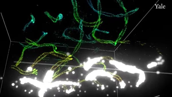
The microscope is able to take detected molecules (below) and resolve them into much clearer structures (above). (Image courtesy of Yale University) |
The Yale University-developed instrument provides researchers with the ability to image entire cells up to about 10 microns in thickness--or about 10 times more than previously possible.
Using a technique similar to the adaptive optics employed in astronomy, the new instrument is able to image the tiny structures inside a cell with remarkable clarity--structures that were previously only observable in small, fractured sections, says Joerg Bewersdorf, associate professor of cell biology and biomedical engineering at Yale and one of the authors on the work.
"Traditional optical microscopes are limited by diffraction to about 250 nm, or a quarter of a micron in resolution," Bewersdorf says. "With our new instrument, we are now able to image whole cells up to about 10 microns, which is about a factor of 10 more than previously possible. We can now see delicate structures at the 10 to 20 nm level in the context of the whole cell, where previous observations were limited to small subsections of a single sample."
http://mdmminn.mddionline.com/?_mc=arti_x_qmedr_edt_aud_parmar_qmedr_med_18_x-Yale3DMicroscope
Bewersdorf says that most optical nanoscopes with some of the best resolution in 3-D use two opposing objectives in a so-called "4Pi" arrangement. Recently two groups demonstrated that 10 to 20 nm resolution is achievable with these nanoscopes, but these instruments are limited in their ability to image sample volumes thicker than one micron. However, this new microscope looks to build off that success, providing images of greater depth.
"Having a resolution of 10 to 20 nm in 3-D now available at our fingertips enables us to study cellular organization close to the size scale of its building blocks, the proteins," Bewersdorf says. "This is an important bridgehead in connecting the structural understanding of individual proteins that we get from structural biology, with the functional observations of traditional live-cell microscopy."
Bewersdorf and his colleagues were able to create the new instrument through the combination of adaptive optics, which corrects for optical distortions that are induced by the sample. He likens this to the adaptive optics in astronomy that can correct for optical distortions of the atmosphere in ground-based telescopes. Then, careful data analysis allows them to pinpoint the locations of individual fluorescent molecules throughout the depth of the sample, providing an image of the structures inside a cell with remarkable depth and clarity.
"Where a traditional light microscope can identify cilia, but cannot resolve ciliary substructures, our microscope can look inside cilia," he says. "This allows us to study the structural implications of ciliopathies, for example. We can see where a particular protein is located at the cilium, and if it is mislocalized in particular genetic disorders."
As the group moves forward with their research, they hope to begin establishing the instrument in labs around the world to provide a tool for other researchers to explore the complexities of cells with this newfound depth and clarity. Bewersdorf remains hopeful that their instrument can provide the means for new discoveries in cell biology and eventually lead to new treatments and therapies for different diseases.
"We are actively collaborating with about five to 10 biological labs at Yale and elsewhere to begin applying our instrument to a diverse set of biological questions," he says. "The first impact we are already seeing now is that this new instrument provides 3-D images of cells at an unprecedented level of detail, which will advance our understanding of the fundamentals of cell biology. As has been proven countless times in the past, this kind of progress in the understanding of the inner workings of a cell will ultimately lead to new treatments for many diseases."
Kristopher Sturgis is a contributor to Qmed.
Like what you're reading? Subscribe to our daily e-newsletter.
About the Author(s)
You May Also Like


.png?width=300&auto=webp&quality=80&disable=upscale)