How 3-D Printing Is Reinventing Benchtop Testing
March 8, 2013
Traditionally, benchtop models, particularly for cardiovascular applications, have been either made of blown glass for rigid parts or silicone for compliant models. Medical device engineers would take the hand-blown pieces of glass or the molded silicone replicas and use it in early product testing. Although the transparency of glass and silicone can be useful, the accuracy of such models is limited and they can be expensive as they are generally made by hand or injection molding.
|
|
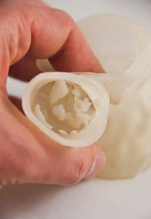
View into calcified aortic valve. 3-D printed from CT scan data with flexible tissue and valve leaflets and rigid calcifications. |
|
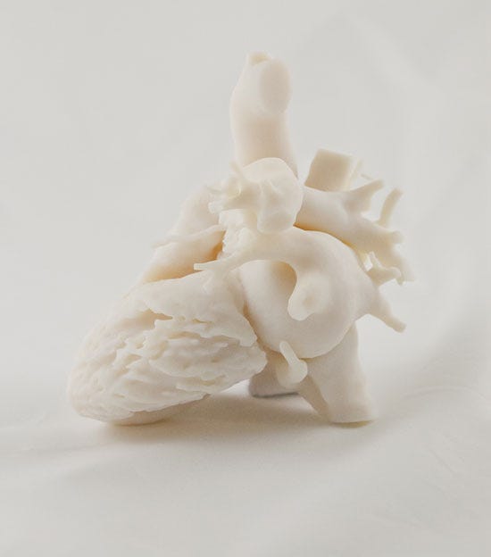
Regulators have recently made recommendations for the use of clinically relevant benchtop models. A recent approach to accomplish that objective is to use 3-D printing to create such models from CT or MRI data. "It allows you to take medical image data and select different regions of the body, whether be soft tissue or bone, and create accurate 3D representations from it," says Peter Verschueren, global cardiovascular business development manager at Materialise (Brussels, Belgium). The company offers a service known as HeartPrint that is used to create such cardiovascular benchtop models. "The methodology enables engineers to quickly identify design flaws that might otherwise not be apparent until later-stage testing such as animal or human trials," Verschueren says. "The 3D printing process helps to minimize a lot of costly trials that tend to happen later in the process by accurately simulating the device deployment up front."
In addition, 3-D printing models can also be used to create representative anatomical models that prove useful in illustrating how medical devices work for specific clinical conditions. For instance, multimaterial anatomical models can be created that include calcifications. "The calcifications are stiff and hard like they would be in the body but the arteries and vessels are malleable," Verschueren says. "It really gives a patient-specific, realistic feel and look to what is going on which is normally almost impossible to model."
Verschueren reports that a number of major players in the cardiovascular market are using 3-D printing technology to create models of patient anatomy--either recreating the anatomy of a specific patient or creating a structure representative of a patient population. The models can be used for device R&D and flow tests as well as intervention training for first-in-man or clinical trials.
|
A 3-D printed model of the heart |
Brian Buntz is the editor-in-chief of MPMN. Follow him on Twitter at @brian_buntz.
About the Author(s)
You May Also Like


.png?width=300&auto=webp&quality=80&disable=upscale)
