How 3-D Bioprinting Is Becoming a Reality
September 7, 2016
Researchers at four universities have been overcoming major obstacles in the field.
Nancy Crotti
|
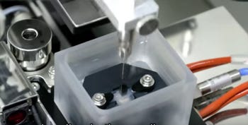
The Regenova 3-D bioprinter loads groups of cells called spheroids into fine needle arrays. (Image courtesy of Cyfuse) |
FDA approval of 3-D-printed body parts may be years off, but obstacles to creating human replacement parts are falling away, thanks to resourceful scientists and new technology.
Researchers at four universities are conquering the barriers, one by one. Indiana University-Purdue University Indianapolis (IUPUI) and Johns Hopkins University are using a bioprinter that preserves cells better than the traditional scaffold 3-D printing technique, according to a report in TechRepublic.
In scaffolding, 3-D printers squirt bio-ink through a nozzle to create objects in layers, a process that can kill the cells. IUPUI and Johns Hopkins are the only U.S. universities that possess a robotic 3-D printer called the Regenova. Made by Tokyo-based Cyfuse Biomedical, Regenova builds structures by placing groups of cells called spheroids in fine needle arrays, according to pre-designed 3D data. The cells can then bond and fuse into tissue.
The Regenova can print tubular body parts, such as veins and arteries, the report said. The IUPUI researchers are working on tissue engineering and regenerative medicine projects in vascular and musculoskeletal biology, dermatology, ophthalmology, and cancer, according to a university statement.
Enabling the cells to create their own external structure--an extracellular matrix--rather than adding it as bio-ink, yields tissues that are much more likely to gain FDA approval, according to Nicanor Moldovan, PhD, an adjunct associate professor of biomedical engineering and of ophthalmology. That's because the gels used in traditional 3-D bioprinting can leave behind fragments that FDA might consider foreign, and even toxic.
"The FDA wants to see the least departure from what is normal in an organism from the standpoint of biosafety and compatibility," Moldovan told TechRepublic. "This doesn't leave a biological signature behind--it's just a way to keep the cells together until they fuse."
Moldovan predicted an FDA-approved construct may be available in at least five years. 3-D bioprinting could prove very lucrative, according to IDTechEx. The British research firm predicts that it could reach an almost $9 billion market by 2014.
At Wake Forest Baptist Medical Center in Winston-Salem, NC, researchers printed ear, bone, and muscle tissue that matured to replace injured or diseased tissue of any shape, according to their study reported in Nature Biotechnology.
The scientists used an integrated tissue and organ printing system that deposits biodegradable, plastic-like materials to form the shapes of ears, bones, and muscles, along with cells contained in hydrogel, TechRepublic reported. The researchers believe these structures could one day be used in humans.
At Penn State, researchers have jumped from 3-D printing artificial cow cartilage in June to experimenting with human cells. Cartilage has only one cell type and no blood vessels. The researchers made cartilage tissue strands and fused them to form a single piece of cartilage.--and they have already begun experimenting with human cells.
A company called Advanced Solutions Life Sciences is working with the University of Louisville (KY) to build a 3-D heart. The team is building capillary beds to enable blood flow with plans to add different cell types, such as heart and liver.
Nancy Crotti is a contributor to Qmed.
Like what you're reading? Subscribe to our daily e-newsletter.
About the Author(s)
You May Also Like


.png?width=300&auto=webp&quality=80&disable=upscale)
