3-D Mammography Could Improve Breast Cancer Screening
June 27, 2014
|
|
Early in June, we reported that General Electric's Healthcare division had received FDA approval for its 3-D Invenia Automated Breast Ultrasound System (ABUS), a next-generation ultrasound breast imaging technology said to detect a third more cancers in women with dense breasts than mammograms alone.
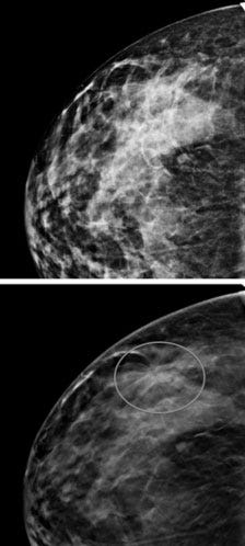
Now, in an analysis of nearly half a million women published in the June 25 issue of the Journal of the American Medical Association (JAMA), researchers have found that 3-D x-ray mammography, or tomosynthesis, combined with traditional x-ray screening, was linked to a 41 percent increase in the detection of invasive cancers as well as a 15 percent drop in the recall rate. This means that fewer women were brought back for additional screening due to false positives.
The study that resulted in the JAMA article, "Breast Cancer Screening Using Tomosynthesis in Combination With Digital Mammography," (Friedewald, et al.) was conducted at 13 academic and nonacademic breast centers from 2010 to 2012. The authors say the combined mode of digital mammography plus tomosynthesis approved by the FDA "addresses the primary limitations of conventional screening mammography by increasing conspicuity of invasive cancers while concomitantly reducing false-positive results."
The authors went on to say that "(t)otal radiation dose when tomosynthesis is added is approximately 2 times the current digital mammography dose but remains well below the limits defined by the FDA," and concluded, "(The a)ddition of tomosynthesis to digital mammography was associated with a decrease in recall rate and an increase in cancer detection rate. Further studies are needed to assess the relationship to clinical outcomes."
In traditional 2-D mammography, the radiologist takes side-to-side and top-to-bottom images of the breast. Tomosynthesis takes multiple x-ray pictures of each breast from many angles. The x-ray tube moves in an arc around the breast while a number of images are taken during the examination. That digital information is sent to a computer, where it is assembled to produce clear, highly focused 3-D images that can "look through" the breast.
While it is not yet clear which modality, tomosynthesis or 3-D ultrasound, has the advantage - and indeed that answer could be different for different patients - it does appear that both of the new 3-D technologies offer a significant improvement over traditional 2-D mammography.
Refresh your medical device industry knowledge at MEDevice San Diego, September 10-11, 2014. |
Stephen Levy is a contributor to Qmed and MPMN.
About the Author(s)
You May Also Like


.png?width=300&auto=webp&quality=80&disable=upscale)
