Sign up for the QMED & MD+DI Daily newsletter.
How Synthetic Diamond Could Revolutionize MRI
Brian Buntz
June 10, 2013
3 Min Read
.svg?width=850&auto=webp&quality=95&format=jpg&disable=upscale "How Synthetic Diamond Could Revolutionize MRI")
Advances in magnetic imaging have contributed immensely to medical science. From the brain to the heart, the bodies of humans and animals generate weak magnetic fields that techniques such as magnetic resonance imaging (MRI) measure to aid in illness diagnosis and treatment. While these techniques have revolutionized medical imaging they are limited in the spatial resolution they can achieve due to the sensitivity of the detectors. However, techniques to image processes on the cellular level will enable a new revolution in medical science, for example, by allowing the study of protein structure or for detailed imaging of a living cell. (Ed note: a blog post titled "Radically Boosting MRI Sensitivity with Hyperpolarized Diamond Nuclei" provides an example of a breakthrough in this vein.)
|
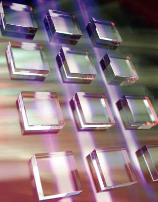
Single crystal synthetic diamond made through chemical vapor deposition. Image from Element Six. |
Achieving such a minute level or resolution calls for the ability to image magnetic fields on the nanoscale, requiring a magnetometer that can measure 3 nT at a distance of 10 nm--a formidable task. Recent progress has been made towards this goal using quantum assisted techniques to manipulate a specific defect in diamond, the so called nitrogen vacancy (NV) defect. Synthetic diamond made using chemical vapor deposition techniques (CVD) allows the production of tailored diamond by manipulating the defects that are incorporated during growth.
These NV defects have a particular quantum property called spin, which is responsible for magnetism and, using simple techniques, the spin of a single defect in the crystal lattice can be used to measure the local magnetic field. A range of research groups have recently used synthetic diamond from Element Six as the basis material to push the boundaries towards cellular MRI (see for example Science 339, 557 (2013) and Science 339, 561 (2013) and Nature 496, 486 (2013)). In their research they have shown that the NV center senses nanotesla field fluctuations from protons with sufficient resolution to measure as few as 10,000 protons in a volume of only 125 cubic nanometers, which approaches the level of individual protein molecules.
Low-Field MRI Systems
In addition to using diamond to sense processes on the cellular level, it may even be possible to use diamond's high magnetic field sensitivity to create new types of low-field MRI systems to replace conventional systems used in hospitals. In a low field, MRI smaller magnetics are required to enable the MRI signal to be recorded due to the higher sensitivity of the detectors used. If this is possible, it will have profound implications for medical imaging by reducing the capital and running costs and increasing the size of volume that can be measured, which is an issue for the obesity problems the western world is currently facing.
While a lot of work still needs to be done to enable diamond MRI systems, the progress that has been made in such a short space of time has put NV centers in diamond on the roadmap as a technique to substantially enhance our understanding of biological processes as well as many additional applications.
Daniel Twitchen is the chief technology officer of Element Six, an international maker of synthetic diamond.
About the Author(s)
You May Also Like

.png?width=300&auto=webp&quality=80&disable=upscale)