Sign up for the QMED & MD+DI Daily newsletter.
A Closer Look at Next-Gen Imaging
Brian Buntz
March 6, 2013
9 Min Read
.svg?width=850&auto=webp&quality=95&format=jpg&disable=upscale "A Closer Look at Next-Gen Imaging")
Researchers from Boston to Austria are working on projects that are paving the way for the future of medical imaging.
After the German physicist Wilhelm Röntgen discovered x-rays in the late 1800s, imaging technology began to have a profound influence on medicine. The past decades have continued to see important developments, including the debut of such imaging modalities as sonography, computed tomography (CT), and magnetic resonance imaging (MRI).
Recently, the role of diagnostic imaging has continued to expand, fueled by advances in computing and other technological breakthroughs. For instance, the rapid acceleration in computing horsepower has made it possible to create sophisticated 3-D reconstructions based on slices from imaging technologies such as CT and MRI. 4-D imaging is becoming more common as well, enabling clinicians, for instance, to use CT to observe the beating of the heart. Imaging software can also be used to optimize CT scans, maximizing their clarity while minimizing the radiation dose required to create them.
While established medical device companies are tapping the ever-increasing power of computing to give imaging technologies new and expanded functionalities, researchers across the globe are exploring the development of completely new imaging breakthroughs that could revolutionize the field in years to come.
Super-High-Speed Imaging
One example of such a development is a camera that can view up to one trillion frames per second. Developed by the Media Lab at the Massachusetts Institute of Technology (MIT; Cambridge, MA; www.mit.edu), the imaging technology is so fast that it can be used to create a slow-motion video of laser light pulses. Last year, the group demonstrated that functionality with slow-motion footage of a pulse slowly oozing from one end of a Coke bottle to the opposite end. In each frame of the footage, light moved less than one millimeter. "There's nothing in the universe that looks fast to this camera," quips one of its developers, Andreas Velten.
Dubbed 'femto photography' by the researchers, this imaging modality can reveal information our eyes normally miss. Its ability to capture the world at such a rapid clip also gives it another unique ability: It can see around corners. This feat is possible by analyzing the trajectories of pulsed light as it bounces off objects. Using that information, the researchers can create models of the objects the light has touched before bouncing off of them. In one experiment, the camera revealed a wooden stick figure hiding behind a miniature door. The camera achieved a submillimeter level of depth precision while viewing a hidden cubic space measuring 40 cm3.
|
Capable of seeing around corners, MIT's super-high-speed imaging technology could potentially be used in a range of applications, including cardiology, gastroenterology, and pulmonary medicine. |
In experiments like this, a series of laser pulses are directed at a visible target. Some of the photons in the laser packets scatter, eventually reaching areas that are invisible from the camera's line of sight. A tiny fraction of those photons bounces back and returns to the camera. "The key is that they reach the camera at a slightly different time slot," Ramesh explains. "Because the camera can sample the world at a trillion frames per second, we can analyze these photons that arrive slightly later. We will piece that information together, very much like you would in a CT scan machine."
|
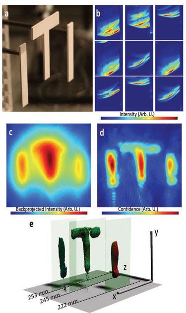
The above images demonstrate the depth and lateral resolution of MIT's imaging system. Figure a shows three letters, which are hidden from the camera's line of sight. Figure b shows images of the letters collected using the camera. Figure c depicts the projection of the heatmap on the x-y plane created by the back projection algorithm. In figure d, the image has been filtered. The color in these images represents the confidence of finding an object at the pixel position. Figure e shows a rendering of the reconstructed 3-D shape. |
Ultimately, this technology could be used to create "diagnostic visualization tools that are not really invasive," Ramesh says. The modality could also be integrated with MRI and CT imaging. One of the technology's most promising applications is endoscopy. Endoscopes that can peer around hidden spaces could prove especially useful when imaging irregular surfaces within the heart or locating hidden polyps in the colon.
While there has been a steady trend toward miniaturization of endoscopes, it is difficult to make them smaller than approximately a millimeter in diameter because of limitations related to their resolution and imaging clarity. While digital endoscopes have image sensors placed at their tip, there is naturally a limit to their reach inside of the body. When used in endoscopes, MIT's imaging technology could enable endoscopes to peer deep inside the body while traveling relatively small distances within it. In addition, the technology could be used externally to peer beneath the skin, working in a manner similar to x-rays but with visible light. Used for this purpose, the technology could potentially be employed in surgical robotics, in which it could enable surgeons to view the insides of body cavities rather than merely the surfaces directly in front of them.
At present, these ultrafast imaging devices are bulky, and the researchers are therefore exploring applications in which portability is not an issue. For endoscope applications, the team is using coupled fibers for imaging and illumination. Ultimately, research on femtosecond solid-state lasers could enable the miniaturization of future-generation technologies by simplifying the camera's illumination mechanism.
Unsurprisingly, the camera itself is a highly complicated piece of equipment. It makes use of 672 optical sensors, a femtosecond laser illumination system, and picosecond-accurate detectors. A titanium sapphire laser that emits pulses roughly every 13 nanoseconds serves as the light source. Sophisticated mathematical reconstruction techniques are employed to enable the instrument to see around bent trajectories. Also, ultrafast video footage results in large files. One second of recording at that speed generates roughly one petabyte of data.
Nanoscale Resolution
While the MIT research is notable because of the blazing speed at which it can capture images, researchers at the Institute of Photonic Sciences (ICFO; Castelldefels-Barcelona, Spain; www.icfo.eu), Consejo Superior de Investigaciones Científicas (CSIC; Madrid, Spain; www.csic.es), and Macquarie University (Sydney, Australia; mq.edu.au) are exploring an imaging modality that is incredibly precise. The imaging modality is similar to MRI but offers a resolution roughly one-million-fold greater.
Ultimately, the imaging modality could be used to facilitate the early detection of conditions such as cancer, improving the patient's likely outcome in the process. The research also opens up the possibility of analyzing the magnetic resonance of isolated cells, which could provide a new avenue for noninvasive disease diagnosis.
Normal MRI works be detecting magnetic fields of atomic nuclei within the body that have been excited by a strong electromagnetic field. While conventional MRI is widely used to image the heart, muscles, the brain, and certain cancers, its diagnostic resolution is limited to the millimeter scale.
In contrast, the scientists at ICFO, CSIC, and Macquarie University, led by ICFO professor Romain Quidant, have developed an imaging technology that boasts a resolution at the nanometer scale, which is sufficient to detect weak magnetic fields from proteins. However, unlike a previous imaging technology that achieved similarly high resolution in the lab using individual atoms at a near-absolute-zero temperature, the new technology enables ultrahigh resolution to be achieved at room temperature. The researchers accomplished this feat by using doped diamond nanoparticles and artificial atoms. And unlike the previous high-resolution imaging modality, which cannot be used in clinical applications, the new breakthrough could enable the development of a novel ultra-high-resolution, noninvasive diagnostic method that sheds light on the inner workings of individual cells.
To manipulate the nanodiamonds, the researchers used laser light as optical tweezers. The tweezers enabled the researchers to manipulate individual nanodiamonds, potentially enabling them to be used as building blocks for high-resolution sensing applications as well as for quantum optics and spin-based quantum information processing. The artificial atoms were created using a nitrogen impurity captured inside of a tiny diamond crystal. The diamond shell stabilized the nitrogen impurity, clearing the way for the imaging modality to be used in a biological environment.
A Flexible Approach to Imaging
A group of Austrian researchers have developed a new imaging method that is notable for the materials it uses rather than its speed or resolution. For this imaging application, the scientists used a transparent polymer sheet that is flat and flexible. In contrast, the majority of image sensors are rigid, planar, and opaque. In addition, the novel image sensor does not use such integrated microstructures as circuits.
Developed by scientists at Johannes Kepler University (Linz, Austria; www.jku.at), the research was recently summarized in an article in OpticsInfoBase. The thin-film luminescent concentrators (LCs) described in the article are doped with fluorescent dyes that absorb light of a certain wavelength (for instance, blue light), re-emitting it at a longer wavelength (for instance, green light).
In order to make the luminescent concentrator function as an imager, the researchers calculated where light struck across the film's surface by measuring the relative brightness of light reaching the sensor array. Calculating for light attenuation, they were able to measure the relative brightness of light reaching the sensor array.
In the article, Alexander Koppelhuber and Oliver Bimber explain how the grayscale imaging technology could be used in touchless user interfaces that respond to gestures. Such an application could be valuable for surgeons in the operating room, who are frequently unable to use touch screen interfaces because of sterilization concerns. The polymer sheet could also be used as a sensor by wrapping it around objects. Because of the relatively low cost of the polymer film, the sensors could ultimately be disposable.
Koppelhuber points out that luminescent concentrators could possibly be used as artificial receptive fields for building artificial human eyes. "The luminescent concentrator sheet can be brought into any shape (for example, by deep-drawing)," he explains. "It would therefore be possible to mimic the shape of the human eye." "Thus, the luminescent concentrators could be used as an artificial retina that is able to restore peripheral vision."
In addition, the ability of the LCs to form hemispherical image sensors enables the creation of cameras that have a wider field of view with lower aberrations than conventional cameras. "This could help to build smaller endoscopes," Koppelhuber says. "It would also be possible to build a lensless camera (a light field camera) by positioning two LC image sensors behind each other. With this, it would even be possible to remove the lens from an endoscope."
Koppelhuber notes that this camera ultimately has many possible medical device applications but stresses that the LC image sensors are still in an early research phase. While the researchers are not currently working on endoscopic cameras, they may incorporate them in their future work.
About the Author(s)
You May Also Like

.png?width=300&auto=webp&quality=80&disable=upscale)