Active MRI Protocol Illumines Movement in Joints
January 13, 2014
Amazingly detailed images produced by magnetic resonance imaging (MRI) have greatly aided physicians, particularly in orthopedics, in their examinations of patients and diagnoses of their ailments. But due to the time it takes to conduct these scans, they have to date been limited to still images.
|
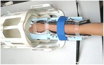
The forearm of a healthy volunteer in the wrist harness is about to be visualized using 'active MRI.' |
Since some joint problems are most apparent during movement of the joint, a University of California, Davis, research team led by Robert Boutin, professor of radiology, has developed a protocol that allows a series of MRI images to be taken in rapid succession, then strung together to create an animation of the joint as it moves. The team's first test results were recently published online in the journal PLOS ONE.
This "Active MRI," as the team has dubbed their development, is "like a live-action movie," says Boutin. "The movie can be slowed, stopped or even reversed as needed. Now patients can reproduce the motion that's bothering them while they're inside the scanner, and physicians can assess how the wrist is actually working. After all, some patients only have pain or other symptoms with movement."
Although these movies are currently limited to slightly better than two frames per second, Abhijit Chaudhari, senior author and assistant professor of radiology, UC Davis, explains, "Active MRI provides a detailed and 'real time' view of the kinesiology of the wrist in action using a widely available and safe technology."
Thus far, the team has only tried their protocol on healthy volunteers in proof-of-principle tests. Their next step will be "to validate the technology by using it on patients with symptoms of wrist instability," says Chaudhari. "We also want to use Active MRI to study sex distinctions in musculoskeletal conditions, including why women tend to be more susceptible to hand osteoarthritis and carpal tunnel syndrome."
And obviously wrists are only the beginning. As the technology is advanced, doubtless frame rates will improve and the range of joints that can be imaged will expand. The eventual impact on sports medicine alone is likely to be huge, and kinesthesiologists everywhere should pay attention, as this is likely the wave of their future.
About the Author(s)
You May Also Like


