Sign up for the QMED & MD+DI Daily newsletter.
Analyzing Particulate Matter on Medical Devices
Manufacturers can employ test method strategies to minimize health risks of intravenous medical devices.
14 Min Read
.svg?width=850&auto=webp&quality=95&format=jpg&disable=upscale "Analyzing Particulate Matter on Medical Devices")
TESTING
|
The light obscuration method, shown here, is automated and may be the easiest particle-counting method. |
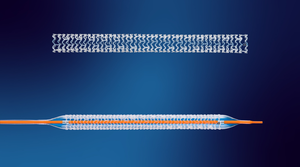
Particulates that may be present on intravenous medical devices such as guidewires, catheters, and stent delivery systems pose potential health risks to patients. When the device is deployed or exposed to the patient, particles may be released and become lodged in the patient's vascular capillary system. Whether this is harmful to the patient depends on the size, shape, and composition of the particles, as well as where and to what degree they cause an occlusion. The response of the patient's immune system to the particles is also a factor.1
The source of these particulates may be the manufacturing process and environment, product packaging, or the device itself. The human body is the dirtiest source in a cleanroom (flecks of deodorant have been found). But clothing, machining, lubricants, and building materials can also be a source of particulates. The integrity of drug coatings found on some stent delivery systems can contribute to the total particulate load. Manufacturers of these types of devices are being asked by FDA to provide data regarding particulates to help evaluate the potential effect the use of their device may have on the patient.
The challenges facing manufacturers include deciding how to test their devices to obtain accurate particulate counts and how to set reasonable acceptance limits for these particulates. Standards addressing these issues are not available for most devices. Acceptance limits for particulates have not yet been established because of the wide range of device types and the unknown effects of particulate size, quantity, and potential toxicity to the patient due to the chemical composition of the particles. Some manufacturers are setting their own acceptance limits or using the limits specified in USP 788, “Particulate Matter in Injections.” However, USP 788 is specific for injections and parenteral infusions and may not always be appropriate for medical devices.
This article evaluates the test methods used to analyze medical devices for particulates, the documentation FDA may request from medical device manufacturers, and the guidelines being developed to help manufacturers establish acceptance criteria for their devices.
Test Methods
USP 788 describes two methods for analyzing particulates in injections and parenteral infusions: light obscuration (method 1) and microscopic (method 2).2 These two methods have been adopted by many manufacturers to analyze particulates on medical devices. For both methods, particles are extracted from the device with particle-free water (i.e., water that has been passed through a 0.22 μm filter) or other appropriate solvent, and the extraction fluid is analyzed. Particle-free water can be easily created in-house with an appropriate filtration apparatus, such as a Millipore jet gun with a filter housing.
|
(click to enlarge) Comparison of the two particulate methods. Light obscuration is the method preferred by USP and should be the default method unless a sample necessitates the microscopic method. |
Light obscuration, which uses laser diffraction technology in a liquid particle counter, is the easiest and most common method used. It is an automated method that determines the size of the particles present in the extraction fluid and the number of each size. A laser diode in the particle counter is directed at the liquid sample, and as particles present in the sample flow through the sensor, they diffract or interrupt the laser diode. This produces a pulse for each particle, and the amplitude of the pulse is proportional to the size of the particle.3
The major advantage of this test method is that, because it is automated, the results are reproducible and nonbiased. The instrument counts and sizes the particles rather than the analyst as in the microscopic method. Sample preparation and analysis is also faster and easier than the microscopic method. The liquid particle counter is capable of counting a wide range of particle sizes from approximately 1.25 µm to 150 µm.4 The exact range varies from instrument to instrument. Another advantage is that the entire sample is not usually consumed in testing, so further analysis can be performed on the extraction fluid if necessary.
However, there are limitations to the light obscuration method. The liquid particle counter is unable to identify particulates by color, shape, or composition. It can only quantitate the particulates and categorize them by size. Any colloid or surfactant present on the medical device may create bubbles when drawn into the sensor of the instrument and be counted as particles. The particle counter may also have difficulty sizing fibrous particulates because of their uneven shape. When such issues arise, the microscopic method is preferred.
|
A particulate with an unusual shape is identified by the microscopic method. But counting the such items can be tedious work. |
The microscopic method is mainly used for samples that cannot be analyzed by the light obscuration method due to turbidity, viscosity, or the presence of colloids or surfactants. The extraction fluid is filtered through a ≤1.0-µm membrane filter. Surfactants and colloids pass through the membrane during filtration, thereby not affecting the results. The filter is allowed to dry, and it is then examined microscopically by the analyst. Particles ≥10 µm are counted separately from those measuring ≥25 µm and sized by means of a circular diameter graticule in the eyepiece of the microscope under 100× magnification. The particulates can also be identified by color, shape, or other parameters. This is helpful in evaluating the possible source of particulate contamination for remedial or preventive measures.
This method also has its limitations. Analyzing samples by the microscopic method is more time-consuming than the light obscuration method. Processing many samples at one time does not provide the typical economies of scale that can be realized with the light obscuration method. Counting particulates on filter slides can be tedious, detail-oriented work. Particles that are clear or the same color as the filter may be missed during analysis. Counts can differ significantly between analysts; therefore, the results are not as reproducible as the automated method. In addition, the entire sample is consumed during the filtration process so further testing is not possible.
|
The microscopic method is used for samples that cannot be tested with light obscuration. It enables partiuclates to be identifed by color and shape. |
When evaluating a device in the laboratory, it is important to ensure that particulates are not introduced into the test system. USP 788 recommends that all testing be performed in an ISO Class 5 hood. All glassware must be washed with a warm detergent solution and rinsed thoroughly with particle free water before being used in the test system.
An area exclusively designed for particulate testing with positive air pressure and HEPA filtration adds an extra level of assurance for minimizing particulate contamination from the laboratory environment. Analysts should be well trained in aseptic technique to further minimize contamination. These are important factors to ensure accuracy and reproducibility and should be considered when evaluating laboratories.
What FDA May Request
FDA looks at what the total particulate load will be to the patient during clinical use of the medical device. The agency is interested in knowing the number and size of particles being shed by the device and where the particles are coming from. It may also want to know the size and number of particulates that are coming from other components used in the test system, such as the guide catheter or guidewire. If the device has components of various sizes, FDA may want all sizes tested for particulates. Some manufacturers have tried to divide their testing process into separate phases to determine what part of the system generates particulates. If the medical device contains components from other manufacturers, it is a good idea to perform baseline particulate counts to determine their effect on the total particulate load of the test system.
A medical device may have a drug or other type of coating that can slough off during deployment of the device, creating particulates. This is especially common among drug-eluting stent systems. If there is a delay between sample preparation and analysis, such particulates may degrade and dissolve, possibly resulting in lower particulate counts than if the sample was analyzed immediately. Therefore, it is recommended that samples of this nature be tested shortly after extraction to minimize the possibility of particulate dissolution.
USP 788 requires that a test environment control be included when analyzing samples by either the light obscuration or microscopic method. This control consists of all items that are part of the laboratory test system, such as the liquid particle counter, glassware, and extraction fluid. The items are rinsed with particle-free water, and the particulate counts of five, 5-ml aliquots are determined. The control must meet the acceptance criteria established in USP 788 before analysis of test samples can proceed.
In addition to the environment control, device manufacturers may want to include a control that is prepared in the same manner as the samples. This control should include all items used in the clinical setting that are separate from the medical device being tested. Examples include guidewires, catheters, hemostasis valves, cannulae, and needles. This control serves as a baseline count of the particulates being shed by components that are separate from the medical device itself.
A common way to evaluate the particulates that the patient may be exposed to during deployment of the medical device is to conduct simulated-use studies with a simulated deployment system. In such a system, devices designed to travel through the bloodstream are subjected to a tortuous path consisting of either a glass or plastic vein that mimics how the device will travel through the patient's vascular system. FDA finds the tortuous path design illustrated in ASTM 2394 to be clinically relevant and recommends that it be used. However, this model may not be applicable for all device systems, so manufacturers may design a similar system and include the rationale for its use when submitting their product for approval. It is advised that the tortuous path incorporate the entire length of the device to which the patient will be exposed. For devices that contain an inflatable balloon, the nominal pressure is routinely used in simulated-use studies. However, for particulate testing, the rated burst pressure may be preferred.
When designing a tortuous path, it is important to include a variety of angles that are representative of clinical usage. However, it is important to avoid creating a simulated-use system that stresses the medical device more than it would be when used clinically. This could result in falsely elevated particulate counts that would be difficult to justify to FDA.
It is recommended that the simulated deployment system be validated prior to actual use, because FDA may request a copy of the validation report. To validate the system, known numbers of National Institute of Standards and Technology (NIST) traceable standard particles of each size used in testing (usually 10 and 25 µm at minimum) are introduced into the tortuous path, and the percent recovery is determined. FDA has established acceptance criteria of a minimum of 90% recovery for 10–25-µm particles and a minimum of 80% recovery for ≥25–µm particles.
The particulate size ranges counted according to the USP 788 method are ≥10 µm and ≥25 µm. Data for larger particulates such as 75 µm, >100 µm, or particulates at the upper limits of the liquid particle counter used in testing are often requested. The presence of large particles on vascular medical devices is a concern because they may have a greater influence on patient health, thereby requiring further justification.
It may also be advantageous to identify the composition of the particulates to determine whether they are homogenous or heterogenous. Knowing the composition distribution of the particulates can provide valuable information about their source, as well as their potential risk to the patient. This is especially important when the counts are high.
Particles of different material types are considered to have a heterogenous distribution. These particles are usually environmental in origin, and their size and numbers are of more importance than their composition. High numbers of particulates made of the same material are termed homogenous and usually indicate a single source. For example, 90% of the total particulate count for a medical device may be due to packaging materials with only 10% coming from the device itself, or there may be a tenfold increase in particulates when a balloon catheter is inflated. A homogenous distribution may point to a specific phase of the manufacturing process as the source of particulate contamination. High numbers of homogenous particles should also be evaluated for their potential toxicity to the patient.
Guidelines
Although there are standards that set acceptable particulate count limits for a few medical devices such as gravity-fed infusion sets (ISO 8536-4), cardiac pacemakers (EN 45502), and autologous transfusion devices (ANSI/AAMI AT6-2005), such standards are not available for the majority of medical device categories. Most vascular medical device manufacturers turn to USP 788 or set their own acceptance limits. USP 788 should be used only as a general guide. Its use is generally acceptable as long as the small volume criteria specified in the method are met. If the particulate counts of the device exceed these limits, an OEM may need to justify to FDA how those counts are acceptable.
An AAMI technical information report (TIR) document that addresses particulates associated with vascular medical devices is currently being drafted. The goal of this document is to provide manufacturers with guidelines for testing and for establishing acceptable particulate levels for vascular medical devices.5 It also focuses on best practices to help manufacturers minimize particulates on their devices through design, development, and manufacturing. The TIR addresses possible sources of particulates from raw materials, the manufacturing process, or the manufacturing environment and provides guidelines for minimizing and controlling contamination. The document is also expected to provide guidelines for manufacturers in setting their own acceptance limits based on such factors as category of the device (i.e., the length of patient exposure to the device) and its clinical application, as well as appropriate particle sizes and their potential toxicity. The author anticipates that specifications will be in counts per device or possibly counts per system is multiple components are used clinically. Specifications may be tied to the category of device. There will likely be an action (warning) limit and an alert limit rather than a simple pass-fail system.
Test methods for sizing and counting particulates are expected to be addressed in the TIR, as should guidelines for sizing particulates by the microscopic method. A list of test methods for analyzing the composition of the particulate contamination and the type of information that can be derived from each method should also be provided in this document.
Conclusion
Many of the recommendations described in this article were gathered through the authors' experience in performing particulate tests and may not apply to all devices. Nevertheless, until a concrete standard is set for particulates on vascular medical devices, it is necessary to consider all FDA recommendations regarding particulate matter testing when submitting a product for approval. Consulting with FDA prior to developing a final draft of a testing protocol can help clarify what data are required for a specific product. A contract laboratory that performs particulate testing can also be a valuable resource for information about what to consider when developing a testing protocol.
Susan Reynolds is a technical writer at Nelson Laboratories (Salt Lake City). Ryan Lunceford is the director for particulate matter studies at the company.
Acknowledgments
The authors thank Tonya Morris, who is on the AAMI Medical Device Particulates Committee responsible for drafting the TIR document.
References
1.LJ Pesko, “Physiological Consequences of Injected Particulates,” in Liquid- and Surface-Borne Particle Measurement Handbook, ed. JZ Knapp, TA Barber, and A Lieberman (New York: Marcel Dekker, 1996): 661–685.
2.USP 788, “Particulate Matter in Injections” (Rockville, MD: United States Pharmacopeial Convention, 2009).
3.HIAC Royco Model 9703 Liquid Particle Counting System Operations Manual (Silver Spring, MD: Pacific Scientific, HIAC Royco Division).
4.Nelson Laboratories internal data, STP0011 Sizing/Counting Particulate Matter.
5.AAMI TIR Draft, “Particulates Associated with Vascular Medical Devices,” (Arlington, VA: Association for the Advancement of Medical Instrumentation).
Copyright ©2009 Medical Device & Diagnostic Industry
About the Author(s)
You May Also Like