Noninvasive Device Scans Skin to Find Tumors
March 1, 2008
R&D DIGEST
|
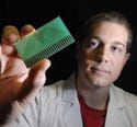
Jye Smith has developed a device that could allow doctors to scan the surface of skin to find tumors. |
A device that scans the skin's surface could identify cancerous tissue without taking a biopsy. By measuring the electrical properties of tissue, the device can tell the difference between healthy and cancerous cells almost immediately.
Developed at Queensland University of Technology (Brisbane, Australia), the system uses bioimpedance spectroscopy to find the boundaries of a lesion via electrical currents.
“[At this time] there are no other bioimpedance mapping devices available,” says Jye Smith, a student finalizing his PhD at the university. Development of the device came out of his research work.
“Bioimpedance has been shown as a useful application before,” says Smith. “However, this device has the beauty of taking multiple measurements over the tissue surface and producing an image so that the operator can see the lesion boundaries clearly.”
By employing impedance measurements at different frequencies, the device determines the electrical properties of the tissue under analysis. The process identifies changes inside the cells and its membranes, as well as in the space between cells. The device, in conjunction with the technique, could diagnose cancers of the skin and cervix.
The prototype uses a device manufactured by Brisbane-based ImpediMed. It has a custom-built multiplexing unit that is attached between an electrode array. The array contains 25 electrodes in a 5 × 5 × 7–mm sheet to allow a surface area of 49 mm2 to be analyzed at a time. It can also be enlarged with more electrodes to increase the measurable surface of the tissue.
“By multiplexing the electrodes around the array, up to 64 measurements can be made at 16 different sites on the surface of the tissue,” says Smith. The sites are displayed as one image to show the areas of varying impedance, which indicate the difference between healthy and cancerous tissue.
Smith says the biggest improvement to the device will be making it handheld. This will enable quick and easy movement to lesions of interest. Other possible changes include increasing the number of electrodes in the array and decreasing their spacing. This will enable larger surface areas to be analyzed, higher sampling rates and image resolution, and clearer identification of lesion boundaries.
The next steps for Smith include finding an industry partner to further develop the device and to continue conducting clinical trials.
Copyright ©2008 Medical Device & Diagnostic Industry
About the Author(s)
You May Also Like


.png?width=300&auto=webp&quality=80&disable=upscale)
