Sign up for the QMED & MD+DI Daily newsletter.
Bob Michaels
January 18, 2011
2 Min Read
.svg?width=850&auto=webp&quality=95&format=jpg&disable=upscale "Using Hydrogels to Grow Blood Vessels in the Lab")
|
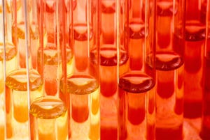
Time-lapse image shows how two types of cells tagged with fluorescent dyes organize themselves into a functioning capillary network within 72 hours. |
Recently, Medtech Pulse reported on research conducted by the National Institute of Standards and Technology and the National Institutes of Health on the use of hydrogels for developing 3-D tissue scaffolds. Hot on the heels of this news, researchers from Rice University (Houston) and Baylor College of Medicine (BCM; Houston) claim to have found a way to employ hydrogels to grow blood vessels and capillaries.
As detailed in the journal Acta Biomaterialia, a team of researchers led by Rice University bioengineering professor Jennifer West and BCM molecular physiologist Mary Dickinson modified polyethylene glycol (PEG), a nontoxic plastic widely used in medical devices and food, to mimic the body's extracellular matrix-- the network of proteins and polysaccharides that make up a substantial portion of most tissues. The scientists combined the modified PEG with two kinds of cells, both of which are needed for blood-vessel formation.
Using light that locks the PEG polymer strands into a solid gel, they created soft hydrogels containing living cells and growth factors. Then, they filmed the hydrogels for 72 hours. By tagging each type of cell with a different colored fluorescent marker, the team was able to watch as the cells gradually formed capillaries throughout the soft, plastic gel. (See video below.)
"The inability to grow blood-vessel networks--or vasculature--in lab-grown tissues is the leading problem in regenerative medicine today," West remarks. "If you don't have blood supply, you cannot make a tissue structure that is thicker than a couple hundred microns."
To test the vascular networks, the team implanted the hydrogels into the corneas of mice, where no natural vasculature exists. After injecting a dye into the mice's bloodstream, the researchers confirmed that there was normal blood flow in the newly grown capillaries.
Associated with this breakthrough, West and colleagues have developed a new technique called 'two-photon lithography, an ultrasensitive method for using light to create intricate 3-D patterns within soft PEG hydrogels. This patterning technique, West says, allows the engineers to exert a fine level of control over where cells move and grow. In follow-up experiments in collaboration with the Dickinson lab at BCM, West and her team plan to use the technique to grow blood vessels in predetermined patterns.
About the Author(s)
You May Also Like