Swallowed Capsule Sticks to the Gut
October 1, 2008
R&D DIGEST
|
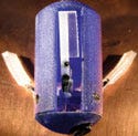
In addition to imaging conditions inside the body, a pill-sized robot is being designed to deliver drugs to targeted areas. |
Pill-sized imaging devices have been designed before. But now one in the works not only is an imaging device, it could also be a vehicle for treatment.
A significant problem with today's camera pills is that they can only retrieve and record images; the capsule doesn't control its position in the body, says Metin Sitti, assistant professor at Carnegie Mellon University (Pittsburgh). He is leading a team that has designed a device that uses available imaging technology for the camera but has added position control capabilities to transform the device into a robot.
“Three years ago, we started to work on changing capsules from pure sensors to active robotic devices, because I think it's the kind of technology that, in the near term, can turn into a real product,” says Sitti, who also directs the university's NanoRobotics Lab.
The device can be swallowed and naturally excreted from the body. It measures 14 mm in diameter and is 30 mm long. It has three legs hidden inside the capsule, and when the doctor wants to anchor the device to tissue, the legs are deployed. The opened legs, which have soft footpads, are coated with polymer microstructures and viscous oils, enabling the pads to stick to the surface of the tissue.
“They can apply good friction, so the device will stop and not move at that given time,” says Sitti. “The three legs are like an umbrella opening and closing.” When the capsule must change its position, the legs are closed, and the peristaltic motion of the intestines moves the device.
When developing the adhesive aspect of the device, Sitti's team examined an insect model. The foot hairs of beetles release an oily substance that helps them adhere to surfaces. The adhesive created by the Carnegie Mellon researchers works in a similar manner. The adhesive is able to secure the capsule inside the body without tearing tissue, and it can attach and reattach. It is also gentle enough that it should be able to safely stick to the intestines, stomach, heart, esophagus, and kidneys.
After the team is able to anchor the device in a desired location, Sitti wants to start looking into more-advanced applications such as removing cancer cells, applying heat, or delivering drugs. This could enable the device to treat diseases rather than simply capturing data on health conditions. The device should be able to operate with commercially available imaging software.
|
Sitti says the next step is to figure out how to enable doctors to control the device. The team needs to develop its own interface and communication network so that the capsule can be remotely anchored and wirelessly operated according to a clinician's needs.
Power consumption is also critical. Small micromotors control the opening and closing actions of the legs, and the researchers are using battery power to activate it. Eventually, the device will be a battery-integrated, robotic capsule. Sitti is also confident that his team will be able to make the device smaller for commercial use by optimizing its manufacturing process.
In addition to scaling the size of the device, the researchers plan to conduct in vivo animal testing to show the feasibility of the robotic capsule. “The final step is integrating tools such as biopsy, heat treatments, or drug-delivery devices on the capsule,” says Sitti. “We're designing them, but we can't integrate them into the capsule robot yet.”
Copyright ©2008 Medical Device & Diagnostic Industry
About the Author(s)
You May Also Like


