Cancer Test Scans Surface Blood Vessels
January 1, 2008
R&D DIGEST
|
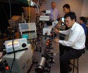
The Purdue team hopes a device firm will create a low-cost microscope for use in |
A minimally invasive test for detecting tumor cells that doesn't require drawing blood could be a faster and more sensitive approach to cancer screening. But researchers at Purdue University (West Lafayette, IN) need a device company to create a low-cost microscope that scans surface veins to identify cancerous cells.
Purdue researchers developed special dyes that selectively attach to cancer cells. Once a fluorescent dye is placed on low-molecular-weight, high-affinity ligands, the conjugate is injected into the bloodstream, where it binds to any circulating tumor cells. Instead of taking a 10-ml blood sample, doctors would be able to put a device up to a patient's skin, and the device would count the dyed tumor cells to assess a large volume of blood in just 20 minutes. A computer could provide a reading on how many tumor cells flowed through the vessel during that period of time.
The current multiphoton microscope that the researchers are using to identify cancerous cells is far too expensive for use in a clinical setting. “One doesn't need one of these microscopes with all the bells, whistles, and additional functions on it,” says Philip Low, professor of chemistry at Purdue. “Doctors aren't going to pay half a million dollars. We'd like to see someone come in and design either a confocal or multiphoton microscope that can be dedicated exclusively for this application, with a price range between $20,000 and $30,000.”
Low foresees using inexpensive multiphoton or single-photon standard linear optical equipment. A handheld laser pointer would provide a long enough wavelength to excite the dyes. Confocal optics would hook onto the microscope and focus on a near-surface blood vessel. A rapid laser scanner would scan back and forth across the vessel, at a rate of about 500 times per second.
Such a device does not yet exist. “We would like to find someone or a company with the capabilities to build and test this,” says Low. “I think the next step is to meet with optical engineers, build the device, test it in animal models, and then take it to clinical trial. I've already had strong interest in testing [the method] from several places.”
The technology would enable immediate identification of malignant cells. According to Low, spiking experiments have shown that the test is extremely sensitive, because it can see every single cancer cell. When the researchers added a specific number of cancer cells to animals, the test detected each one. The test could also be used immediately after a patient has chemotherapy to identify any residual disease. This could have a huge effect on ovarian cancer, for example, because by the time recurrence is diagnosed, it's often too late.
“A malignant mass sheds about 1 million cancer cells per gram of malignant mass per day, so we should be able to pick that up very easily,” says Low. “Rather than wait 14 months to decide whether the patient should have more-aggressive chemotherapy, we could start it immediately because we would still see circulating tumor cells.”
The Purdue researchers have also been working with the Mayo Clinic on the technology. Their work has been funded by an Indiana Elks Charities grant, the Purdue Cancer Center, and an Ovar'coming Together (Indianapolis) research grant.
Copyright ©2008 Medical Device & Diagnostic Industry
About the Author(s)
You May Also Like


