Implant Restores Inner Ear Balance
October 1, 2007
R&D DIGEST
|
Charles Della Santina of the Johns Hopkins University School of Medicine says a 3-D prosthesis is key to helping patients regain inner-ear sensory cells for balance. |
Johns Hopkins researchers are developing a device similar to a cochlear implant that partially restores balance in patients who have lost crucial sensory cells. The multichannel prosthesis senses head movements and sends the information to the brain via electrodes connected to the vestibular nerve.
“Our new device measures and encodes head rotation in all three dimensions, stimulating three or more branches of the vestibular nerve,” says Charles Della Santina, MD, PhD, assistant professor of otolaryngology at the Johns Hopkins University School of Medicine (Baltimore). “Because we move our heads in a 3-D world, this is an important step toward a clinically useful prosthesis for people who have lost their inner-ear sensory cells for balance.” Della Santina is also director of the Vestibular Neuroengineering Laboratory at Johns Hopkins.
Inner-ear hair cells are the sensory receptors for balance. The loss of that function can happen when patients are required to take high doses of gentamicin, an antibiotic used to treat serious bacterial infections such as meningitis or Ménière's disease. A side effect of the medication is that it can wipe out those cells. Other illnesses and viral infections can result in a loss of balance as well.
The Johns Hopkins team demonstrated the implant's proof of concept on rodents. The current device is enclosed in a plastic case about the size of a small dental floss container, with eight electrode wires coming out from one side. Electrode leads pass through an acrylic percutaneous connection to the inner ear.
|
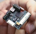
The vestibular implant circuitry is still too large and thick to be implanted into humans, but Johns Hopkins researchers have shown proof of principle. |
The sensors, which are connected to the electrodes and a microprocessor, measure the speed at which the head rotates. After the information is processed, the device measures the time it takes electronic pulses to travel through the electrodes located near the branches of the vestibular nerve that react to head rotation. In the rodent study, the pulses lasted less than a millisecond.
For use in humans, the device must be thin enough to fit into a small depression in the skull without protruding from beneath the skin. The device will probably be about 1 × 1 in. long and 6 mm thick. The researchers are working on using smaller and thinner 3-D rotation sensors to meet the low-profile requirement.
Other changes include reducing power to extend battery life and implementing a transcutaneous power and control link similar to cochlear implants. The team also needs to refine the electrode design to maximize its ability to selectively stimulate each branch of the vestibular nerve without creating crosstalk to the other branches of the nerve, Della Santina says.
Because the device's housing materials will be similar to those used in cochlear implants, Della Santina doesn't foresee any biocompatibility issues. The prosthesis will use Teflon-coated, platinum-iridium wires encapsulated in silicone. A hermetic metal canister will house the circuitry.
A patent is pending on certain parts of the implant. The Johns Hopkins study of the device was funded by the American Otological Society and the National Institute of Deafness and Other Communication Disorders, a member of the National Institutes of Health. A report on the prosthesis was published in the June edition of IEEE Transactions on Biomedical Engineering.
Copyright ©2007 Medical Device & Diagnostic Industry
About the Author(s)
You May Also Like


.png?width=300&auto=webp&quality=80&disable=upscale)
