Sign up for the QMED & MD+DI Daily newsletter.
Growth-Factor Coating Shown to Improve Healing of Bones Around Implants
mkingsley
February 8, 2012
3 Min Read
.svg?width=850&auto=webp&quality=95&format=jpg&disable=upscale "Growth-Factor Coating Shown to Improve Healing of Bones Around Implants")
|
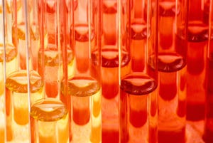
An experimental growth-factor method developed by University of Wisconsin-Madison biomedical engineer William Murphy for attaching implants to bones produced strong, healthy bone as the bone healed around a metal implant in an animal model (right). The implant on left was performed with conventional surgery techniques and produced less bone and more scar tissue (white areas). Implants are the large black areas. Image: University of Wisconsin-Madison/nanowerk.com |
Orthopedic implants dip-coated with modular growth factors can stimulate bone and blood vessel growth in sheep, two researchers reported at the Orthopedic Research Society meeting in San Francisco earlier this week. The researchers, Darilis Suarez-Gonzalez and Jae Sung Lee, are from professor William Murphy's biomedical engineering lab at the University of Wisconsin-Madison.
Growth factors--proteins that can stimulate tissues to grow--have posed a challenge to medical scientists in that the proteins can be too effective or not specific enough, leading to cancer rather than the controlled growth needed for healing. Murphy wants to start applying the manifold benefits of a modular approach to healing or regenerating bone, tendon, and ligaments. In particular, he hopes to leverage this method in replacement surgery after an artificial joint has loosened or failed. Temporarily stimulating bones to grow by placing growth factors near the new implant could shorten healing time and ensure a good, tight fit.
With his approach, the working end of the modular structure may feature a fragment of a growth factor, but not the entire protein. "Often, you just want the specific regions that activate the signaling pathways, because that can reduce the chances of stimulating unwanted growth, even cancer," Murphy says. At the other end, Murphy may place an anchoring molecule that binds to the bone and prevents the modular structure from migrating away from the wound.
With the modular approach, he says, "you might be able to stimulate bone formation without the side effects. We are trying to decrease stimulation outside of the bone defect, trying to design these molecules to specifically generate new bone in a defect, and to stay there." Animal tests of the modular technique, performed in collaboration with Mark Markel, a professor of veterinary medicine, have shown that the bone is denser around the implant, and that the union between the implant and the bone is stronger than those produced by state-of-the-art orthopedic techniques. The added growth factors have not been detected elsewhere in the animal, Murphy says.
The approach could also be used for reattaching ligaments to bone after sports injuries and healing large bone defects during spinal fusion, facial reconstruction, or trauma. For this type of work, Murphy collaborates with two associate professors of orthopedics and rehabilitation at the School of Medicine and Public Health. "Ben Graf focuses on knee injuries in sports medicine," he says, "and David Goodspeed, a lieutenant colonel in the Army who has seen blast injuries during multiple tours in Iraq, is working on the kind of major traumatic wound we think is potentially treatable using this approach."
Moving this technique from the lab to clinical practice is a major step. "We have shown that this can work in a large, clinically relevant animal model; but realistically, I don't see this being used in the clinic within the next five years," Murphy says.
About the Author(s)
You May Also Like