Cell Imaging Faster than the Blink of an Eye
R&D DIGEST
August 1, 2006
|
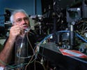
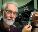
Paul Robinson, PhD, of Purdue University (West Lafayette, IN) has helped design a device that can analyze cell components for irregularities in a split second. |
A method that produces detailed images of Earth is being applied in diagnostics to identify the structure of cells and tissues. The technology reveals information quickly and could become an economical way to identify diseases.
Researchers at Purdue University (West Lafayette, IN) have adapted the functions of a Landset satellite. First, the satellite flies around and takes images of the land. Then, using sophisticated algorithms, it unmixes complex spectral information that can reveal, for example, wheat and barley, along with any diseases the crops have. “Here, you're looking from a remote perspective and making very sophisticated analyses,” says Paul Robinson, PhD, professor of cytomics and director of Purdue's cytometry laboratories. “We're looking at the same thing from a proximal perspective.”
The technique can rapidly analyze separate colors in cells or particles. Robinson's group uses a modification of flow cytometry, a method in which cells are placed in liquid and analyzed as they flow past a laser beam. “A regular flow cytometer has reached its limit,” says Robinson. “This opens it up more for clinical diagnostic use.”
With the new method, researchers can look at blood cells, tissue cultures, and bacteria, and analyze the innate components of the cells. They can also add dyes and labels to the cells to code them, which Robinson calls spectral bar coding. The technology enables the researchers to see the complete spectra of a cell as it appears in front of the laser beam, which is only for about 0.001 second. A complete fluorescence profile of the cell, including any abnormal characteristics, is made, literally, faster than the blink of an eye.
|
Robinson hopes the device, which is already smaller than a conventional flow cytometer, can be reduced to a cubic foot or smaller. |
“I think if you look at where flow cytometry is inside clinical diagnostics right now, it doesn't cover a great range of areas,” says Robinson. “We have advanced classification algorithms and sophisticated sets of data, so there will be opportunities to use that hardware and software integration to apply to other areas that we haven't even thought about.” For example, microbiologists could use the technology to classify microorganisms, he says.
It could also be applied to diagnostics for a range of diseases. “We should be able to take the white or red blood cells and extract an enormous amount of information from them and identify how [cell components] fit with known diseases,” says Robinson.
Although not at its ideal size—the device should be about a cubic foot or smaller—Robinson says all the necessary parts have been integrated. Getting the device to a compact size will be the job of the company that commercializes it, he adds. “The [parts] have to become smaller, cheaper, and more efficient before [the technique moves] into the medical arena.”
The size of the unit is already smaller than a conventional flow cytometer. Instead of dozens of optics, it has only one or two. Components include a detection system, electronics, microfluidics, multiple lasers, and software. According to Robinson, the electronics are more sophisticated than most flow cytometers today, which are only capable of 8–10 channels of data collection. The new technology that Purdue is working with uses 64 channels on one board. A small company developed this aspect of the device for a military application. “We're not trying to reinvent every part of this new vehicle. We're using the advances in technology that are naturally occurring in industry,” says Robinson.
The device will likely evolve with advances in nanotechnology and microfluidic technology. “We'll have opportunities for embedding some of our analytical and detection tools in microfluidic technology where tiny volumes of liquids are used, as opposed to the milliliters that we use today for clinical blood evaluation.”
The last part of the system that will be updated is the software and data analysis, which is becoming quite sophisticated. “It requires advanced mathematical modeling and sophisticated algorithms that can do high-speed calculations, evaluate mixed data sets, and look for pattern recognition,” says Robinson.
He adds that the ability to do classifications in real time is the holy grail of next-generation analytical instruments. “We've already made leaps into the next generation of software analysis by using very sophisticated classification routines. We can look at 32- to 64-dimensional analysis,” says Robinson. The next step is to make the components operate in an integrated manner and then fit them into a small package.
Copyright ©2006 Medical Device & Diagnostic Industry
About the Author(s)
You May Also Like


