October 18, 2016
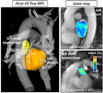
Researchers at Northwestern University have developed a new imaging technique that can image blood flow in three spatial dimensions--helping to identify patients at risk for stroke.
Kristopher Sturgis
 Northwestern University researchers have created a new imaging technique that could help doctors better predict which patients are most at risk for stroke by examining atrial fibrillation and blood flow. Michael Markl, professor of biomedical engineering at the Northwestern University Feinberg School of Medicine and developer of the technology, told university news that atrial fibrillation has been easy to detect, but hasn't really provided any indication of stroke risk.
"Atrial fibrillation is thought to be responsible for 20 to 30% of all strokes in the United States," Markl said. "While atrial fibrillation is easy to detect and diagnose, it's not easy to predict who will suffer a stroke because of it."
Atrial fibrillation has been linked to strokes given its ability to slow blood flow throughout the body, which can lead to blood clots that can travel to the brain to initiate a stroke. Markl and his colleagues began their study to develop a cardiac magnetic resonance (CMR) imaging test that could detect the velocity at which the blood flows through the heart and body.
This technique, known as atrial 4-D flow CMR, was designed to provide a non-invasive approach that comes in the form of software, making it easy to integrate into current MRI equipment without any need for special equipment, scanners, or hardware. Markl says that the new technique was essentially a process of retooling the scanner to process information in a new way.
"We simply programmed the scanner to generate information differently, in a way that wasn't previously available," he said. "It allows you to measure flow, diffusion of molecules, and tissue elasticity. You can interrogate the human body in a very detailed manner."
Recent advancements in MRI imaging techniques have begun to bear fruit, as researchers continue to look for new ways to see inside the body. Earlier this year a British research institute introduced "salt" MRI scans that can identify sodium concentrations in the body to help detect disease. If that's not wild enough, researchers from the University of Nottingham in the UK aim to enhance lung imaging through the use of krypton gas as a contrast agent to reveal new areas of the lung on an MRI scan.
These recent advancements look to evolve traditional MRI imaging techniques to help doctors visualize new areas of the body for diagnosis and treatment. When it comes to Markl's new technique, the aim is to better identify which atrial fibrillation patients are at high risk for stroke, so they can begin treatment to prevent blood clots. These treatments are effective at reducing the risk of stroke, but they can also increase the risk of bleeding complications -- making it crucial to identify which patients are truly at risk for stroke.
In the past, doctors have attempted to assess stroke risk in atrial fibrillation patients through the use of a risk scoring system that accounts for risk factors like age, gender, and general health. Markl designed the new 4-D flow imaging technique to provide doctors with a tool to better assess stroke risk based on actual blood flow dynamics, which should more accurately identify high stroke risk and prevent over treating patients.
As the group moves forward with their research, Markl plans to continue to observe atrial fibrillation patients as part of a longterm study in an effort to improve the predictive power and diagnostic abilities of the new technique. He says the plan is to develop and integrate new algorithms and tools that can make it easier and faster to analyze the data and produce results in a matter of minutes -- and when it comes to stroke, a few minutes can make all the difference.
About the Author(s)
You May Also Like


