Sign up for the QMED & MD+DI Daily newsletter.
Nancy Crotti
March 16, 2017
2 Min Read
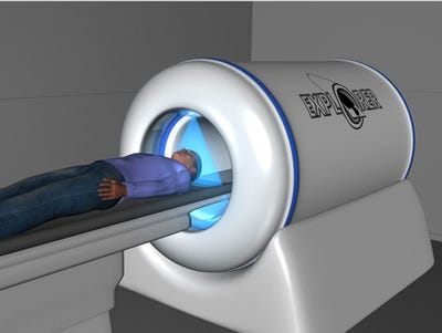
.svg?width=850&auto=webp&quality=95&format=jpg&disable=upscale "Total-body PET, Find Out What it Means to Thee")
Researchers have NIH funding and industry partners for a total-body PET machine that could work faster and produce better images.
Nancy Crotti
U.S. and British researchers are closing in on creating a total-body positron emission tomography (PET) scanner that would work faster and with less radiation to produce higher-resolution images than are currently available.
Led by Simon Cherry and Ramsey Badawi of the University of California, Davis, the team believes that a total-body PET machine could have applications in cancer, immunology, and arterial disease. Faster scans might also aid in treatment of pediatric and adolescent patients, according to Cherry. The team published a paper on its research in Science Translational Medicine.
A PET scanner is used to observe metabolic processes, detecting gamma rays emitted by a radiotracer injected into the bloodstream. Currently, those scans take about 20 minutes and involve small areas of the body, and creating a total-body scan requires multiple imaging sessions.
A total-body PET scanner would be able to capture the entire body at once, eliminating the need for multiple scans. It would also allow for a 40-fold gain in effective sensitivity and a 6-fold increase in signal-to-noise ratio compared with current technology, according to Cherry.
Dubbed the EXPLORER project, the total-body PET scanner research won a $15.5 million grant from the National Institutes of Health in December 2015. The team revealed a mini-prototype one year later, and most recently, enlisted United Imaging Healthcare America, a subsidiary of Shanghai United Imaging Healthcare, and SensL Technologies of Cork, Ireland, to build a full-size prototype.
The biggest callenge has been convincing teh cmt of the value of this idea and getint the funding to actuliy bu ild the first scanner," Cherry said in a phone interview. "The size of teh scanner is roughly the size of eight curent PET scanners put togehr, and PET is already seen as an expsnsive technolgoy. The questionis, will be can we show sufficient clinical benefit to offset the higher cost."
The team envisions initial applications for research, but hopes to make such a scanner clinically available. Such a scanner could cost about $10 million, Cherry noted.
"The benefits here are enormous. Factors of 40 are huge," he said. "As people get interested, there will be huge pressure to bring the costs down. I really think we have a pathway. It will take some time, but ultimately, I think the cost/benefit ratio will be favorable."
Nancy Crotti is a contributor to Qmed.
[Image credit: EXPLORER project]
About the Author(s)
You May Also Like


