The China-based research team foresees a bright future for this novel nanomaterial-based application, especially in clinical and diagnostic setups.
February 24, 2022

Pathology laboratories often need to analyze fluid samples for the presence of dopamine and uric acid. However, the high concentration of ascorbic acid, which is also present in clinical samples, tends to interfere with the accurate determination of dopamine and uric acid levels simultaneously. Now, researchers in China have found a nanotechnology-based fix to this bioanalytical problem, paving the way for efficient detection of dopamine and uric acid in pathological samples.
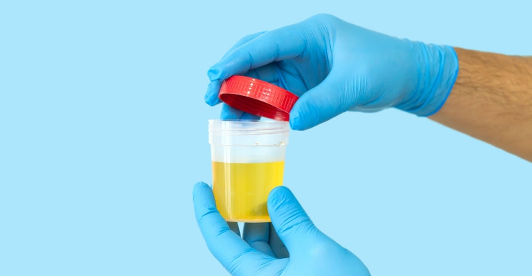
Urinalysis, or the detection and estimation of various pathophysiological substances in urine samples, is routinely recommended by physicians for disease diagnosis. Increased levels of uric acid, for instance, may indicate underlying kidney or heart disease. Similarly, an increase in the urinary levels of dopamine may indicate the presence of neurological disorders like neuroblastoma or Parkinson’s disease. Because pathology labs need to simultaneously determine the urinary levels of multiple substances, techniques that permit such co-detection are necessary. However, such co-analyses sometimes present with technical hurdles. In the case of urine samples, the relatively higher concentrations of ascorbic acid in urine interferes with the simultaneous detection of dopamine and uric acid, both of which are present at relatively lower levels.
A group of researchers led by Associate Professor Dongdong Zhang of Xi'an Jiaotong University, China, has now been able to overcome this limitation through an electroanalytical technique that permits the co-detection of dopamine and uric acid in urine samples even in the presence of absorbic acid.
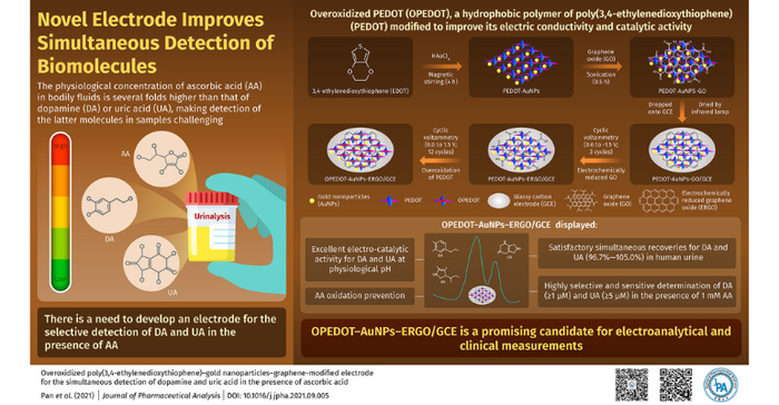
In their paper, published in the Journal of Pharmaceutical Analysis in December 2021, the researchers describe how they combined a nanocomposite mixture, which had an average grain size of 10-9 meters or more, made up of gold nanoparticles (AuNPs), a special (conducting) polymer, and electrochemically-treated graphene oxide over a conventional glassy carbon electrode (GCE), to get a superior electrode for use in their technique. A GCE combines the properties of glass with those of graphite. However, it needed to be modified for the selective and simultaneous detection of dopamine and uric acid in the presence of high concentrations of absorbic acid.
To this end, the researchers employed a combination of chemical and electrochemical methods. They started with poly(3,4-ethylenedioxythiophene), or PEDOT, which is a highly conductive polymer with a lot of promise in the field of biosensors.
“PEDOT can be overoxidized to obtain OPEDOT, whose hydrophilicity and unique properties make it useful in electroanalytical applications. However, since OPEDOT is not as good an electrical conductor or catalyst, it is modified with suitable nanomaterials, in this case, gold nanoparticles," Zhang said.

The PEDOT-AuNPs were chemically synthesized from chloroauric acid and 3,4-ethylenedioxythiophene at room temperature. The addition of graphene oxide (GO) resulted in the formation of a homogeneous suspension of PEDOT-AuNPs-GO. This suspension was then dropped onto the surface of a GCE and dried. Finally, following an electrochemical procedure, the nanomaterial OPEDOT-AuNPs-ERGO/GCE was successfully fabricated and readied for bioanalytical measurements.
When used, the modified electrode was able to simultaneously detect extremely tiny amounts of dopamine (1 mM) and UA (5 mM) under physiological conditions, even in the presence of a large excess (1.0 mM) of absorbic acid.
The research team foresees a bright future for this novel nanomaterial-based application, especially in clinical and diagnostic setups.
“Our novel, graphene-based, ternary composite, with its advantageous features, is a promising candidate for electroanalytical and clinical applications,” Zhang said.
The researchers said future studies are required, but this nanocomposite electrode has strong potential to become the gold standard for diagnostics in pathology laboratories.
About the Author(s)
You May Also Like


