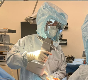
LumaMed, a startup in Georgia, aims to help surgeons detect breast cancer at the margins by a imaging device it has developed.
August 14, 2013

.jpg?width=700&auto=webp&quality=80&disable=upscale)
Repeat breast cancer surgery rates vary widely - sometimes women unnecessarily get them, while some who should undergo another procedure are not operated upon.
That's because the guidelines for when repeat surgeries should be performed are fuzzy at best and largely dependent on a physician's interpretation.
This is the world in which LumaMed aims to play. The Georgia startup wants to put a device that can image cancerous tissue that has been excised after a lumpectomy procedure in real time in the hands of breast cancer surgeons so that they know whether they have removed all the harmful tissue. Or whether they need to go back to the operating table and remove more tissue then and there instead of having to wait to do a second surgery in the future.
Called LumaScan, the device uses "patented technology that provides rapid, intra-operative digital imaging of the surface of tissue excised during cancer surgery."
The device is able to produce images that can distinguish between normal and abnormal tissue as well if not better than conventional histopathology, said Mark Samuels, CEO of LumaMed in a recent interview.
This fall, the company will test a pre-commercial prototype on 40 women who undergo lumpectomies at the University of Wisconsin. The trial is expected to conclude early next year and LumaMed plans to file for FDA clearance in the early summer, 2014.
While the need may be great to have devices that can accurately detect cancer at the edges of tissue, LumaMed is sure to find some competition in the marketplace.
Earlier this year, the FDA approved a novel breast cancer detection device that followed the more rigorous pre-market approval route called the MarginProbe. Developed by Dune Medical, MarginProbe looks at how excised tissue responds to an electric field, thereby creating electromagnetic “signatures” that can be used to identify healthy and cancerous tissue in patients. The technology is based on RF spectroscopy.
Samuels acknowledged that he has been following MarginProbe closely but believes that LumaScan has more powerful technology.
"It sends a RF energy into the tissue and that makes a determination whether or not the conditions indicate that there could be a problem but it doesn't give any kind of picture of exactly where the problem is," Samuels said of MarginProbe. "We think if you are trying to remove breast cancer, or oral cancer - maybe you are trying to remove a tumor that near in the tongue or near the voice box or you are in brain surgery - we think it is very important to have an image that actually shows you exactly where the dividing line [between cancerous and normal tissue] is."
The proof of course will be in the pudding. While MarginProbe has shown that it can reduce repeat lumpectomies by 50% or more, Samuels hopes to learn more about his device's accuracy in the clinical trial at the University of Wisconsin.
So far LumaMed has been privately funded, as well from grants from National Institutes of Health. Samuels hopes to talk to venture capitalists during or after the clinical trial to raise a Series A round. Although the company is aiming the LumaScan at the breast cancer market, the technology can be applied to other cancer surgery as well.
Samuels is also thinking ahead of a next-generation device of LumaScan where the surgeon won't have to wait to examine excised tissue to know whether all the abnormal tissue has been removed. That device would provide tissue visualization to actually help the surgeon decide where to make the cut to get all of the cancerous tissue out.
"Instead of taking the tissue out and then going back in for more, you make that decision correctly from the start," Samuels said. "That is the ultimate goal of LumaMed is to be able to provide a cancer vision that would be able to work in real time and help the surgeon to know exactly where to put the scalpel."
[Photo Credit: iStockphoto.com user ideabug]
-- By Arundhati Parmar, Senior Editor, MD+DI
[email protected]
You May Also Like


