R&D DIGEST
August 1, 2006
|
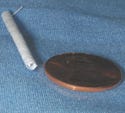
The Bowlin vascular graft shown here measures just 1.5 mm ID. It could one day be used in the hand, ankle, or artery region. |
Designed for vascular grafts, a new material that degrades in the body may also promote tissue regeneration. Although there's hope that the bioabsorbable material could be used for grafts in the coronary artery, success must first be demonstrated in the peripheral arteries. A degradable graft could help treat diabetics at risk for limb loss due to vascular disease and patients with trauma injuries or atherosclerosis. Researchers from Virginia Commonwealth University (VCU; Richmond, VA) reported on the material in the June issue of the journal Biomedical Materials.
The material is designed to be strong and flexible enough to provide support for a graft. Researchers found that a blend of polydioxanone (PDO) and elastin fibers met the criteria.
PDO is a synthetic and biodegradable polymer; elastin is a natural polymer found in the body. “The PDO is fairly nonreactive and it gives us high strength for an extended period of time,” says Gary Bowlin, PhD, associate professor and director of the tissue and cellular engineering lab at VCU.
“The elastin gives us the natural vascular component or protein that can be recognized by the cells and aid in regeneration,” he adds. “So, we get strength and bioactivity.”
The researchers used a technique called electrospinning to create the PDO-elastin blend. Bowlin likens the process to making cotton candy. “All we're doing is taking the polymer solution, the blend of PDO and elastin, and putting it in a reservoir with a nozzle. Then we charge it to a very high voltage opposite some target, which is a tubular structure, some distance away.” When the polymer is charged, an electric field is created in the gap. Then the tubular structure, or mandrel, is grounded. This draws out the polymer solution to form a liquid jet.
“The liquid jet travels through space, the solvent evaporates, and we're left with a nanofiber. That nanofiber then collects on the mandrel to form a nonwoven, nanofibrous tubular structure.”
|
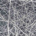
A scanning electron micrograph illustrates the structure of the tube wall. Fibers are less than 500 nm in diameter. Photo courtesy of Scott A. Sell of VCU. |
The structure can be any desired length or diameter. It can also accommodate a tapered design. “A lot of bypass procedures are done in the leg,” says Bowlin. “If you have to go from the groin region to the ankle, you go from 6 to 2 or 3 mm in diameter. We can design the mandrel to provide that taper.”
One of the more popular materials for vascular grafts has been Teflon, but it doesn't degrade in the body and, because it's inert, it is susceptible to infection. Another problem with Teflon is that it's fairly rigid. “Arteries that [the grafts are] adjacent to are fairly elastic, so you have mismatches in terms of mechanical properties and that causes abnormal stress on the cells and abnormal tissue growth that leads to complications,” says Bowlin.
Although the new material isn't as strong as inert materials, it has more-natural characteristics. “We want a compliant structure, something like a rubber band, but we tend to think of those as being weak,” says Bowlin. And polymers alone tend to create more rigid structures. “That's been part of the trade-off in vascular design over the last couple of years. How do we keep the strength but get the elasticity?”
Bowlin says the structure they've created is a nanofibrous blend that has mechanical properties almost identical to a native blood vessel. It should also be strong enough to withstand all arterial forces while simultaneously promoting regeneration as it is absorbed. “Our structure holds a suture better than a saphenous vein, which is a traditional autologous bypass material. PDO and elastin have substantial strength.”
Within a few months to a year, the patient is left with a regenerated artery in place of the absorbed polymeric graft. The material also slowly degrades with fewer adverse reactions than other materials.
However, Bowlin can't pinpoint an exact time frame for degradation, because the researchers haven't developed the final recipe for the PDO-to-elastin ratio. This is currently their biggest design challenge. “The degradation-regeneration timing sequence has to be perfect. If material degrades too quickly, it leads to rupture and failure,” says Bowlin. “If the same foreign or synthetic material stays around too long, the cells may sense it as foreign and not really regenerate into a normal or native structure.”
This is another reason Bowlin wants to avoid the coronary artery for the time being. In the peripheral vascular system, many patients have complications associated with lack of blood delivery to the extremities. For example, the graft would be a last resort in trying to restore blood flow and save a patient's leg. If a rupture or a blockage occurs, the result isn't usually life threatening. However, a rupture or blockage in the coronary area can be catastrophic.
The researchers' work has been supported by a grant from the American Heart Association, Mid-Atlantic Affiliate.
Copyright ©2006 Medical Device & Diagnostic Industry
About the Author(s)
You May Also Like


