Sign up for the QMED & MD+DI Daily newsletter.
Originally Published MDDI June 2006 R&D Digest: The monthly review of new technologies and medical device innovations. Heather Thompson
Heather Thompson
June 1, 2006
4 Min Read
.svg?width=850&auto=webp&quality=95&format=jpg&disable=upscale "Nanofiber Scaffold Supports Optic Nerve Regrowth")
R&D Digest: The monthly review of new technologies and medical device innovations.
|
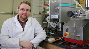
Rutledge Ellis-Behnke, left, and Gerald Schneider created a technique that has helped rodents recover sight after having neural pathways severed. |
Using nanosized peptides, a team of researchers has built knitted scaffolds that may be used to regrow damaged optic nerves. The team from the Massachusetts Institute of Technology (MIT; Cambridge, MA), has managed to restore limited sight to blinded hamsters using nanoparticles resembling small fibers.
To conduct the experiment, the researchers cut the neural pathway that enables vision in hamsters. They then injected 16 of 47 adult hamsters with a solution containing nanofibers. The material was injected into the gap within an hour of the pathway being cut. After the first 24 hours, the researchers noted that the gap was reduced and that axons seemed to have grown through the center of the cut.
When tested, three-quarters of those hamsters could function well enough to identify a food source. None of the 31 hamsters that did not receive the nanosolution regained sight.
According to the lead researcher, Rutledge Ellis-Behnke, the technique offers a possible method for repairing neural connections. When neural pathways in the brain or spinal cord are damaged, they don't usually heal. The damage can result in lifelong brain damage and paralysis.
When a neuron is cut, he says, it sprouts a growth tip, much like a tree whose branch has been cut. After that initial step, however, the growth stalls and scientists are not sure why. Axons can be encouraged to extend by exposing them to growth factors. But they rarely extend far enough to bridge the large gaps typical of most optic nerve injuries, he says.
Ellis-Behnke believes that the nanoparticles may block signals that trigger an immune response. Alternatively, he speculates that perhaps the nanoparticles coat the growing tip of the neuron, blocking any signals that tell the axons not to grow.
Gerald Schneider, one of the team members, estimates that 30,000 axons reconnected in the hamsters, compared with only around 30 in previous experiments using other approaches, such as nerve growth factors. The nanoscaffold is similar in size and shape to sugars and proteins. The team believes that the similarity between the size of the fibers and the features on neural material is what encourages the axons to bridge the gap. The scaffold is biodegradable and appears to eventually break down harmlessly.
And while the results are promising, Schneider explains that the technology is not necessarily a cure-all. “It will not replace neurons that have been destroyed. The axons are slow to grow. We have used the method only for situations in which they have to grow very short distances to get some recovery of functions.”
Schneider says that the scaffold could be included as one of many therapies, such as stem cells or growth factors, to help regenerate nerve connections in people who suffer strokes, spinal cord damage, and brain injuries. “We expect that the method will have to be combined with other treatments in some situations. In humans it will probably be used first in surgery on the brain and spinal cord,” he says.
Still, the scientific community is encouraged by the work at MIT. The knitting “could be very useful in combination with other treatments,” says Wolfram Tetzlaff, associate director of discovery science at the International Collaboration on Repair Discoveries in British Columbia, which focuses on spinal cord injuries.
Tetzlaff cautions, however, that the hamster work involved a clean knife cut across the optic nerve, and “this is not how injuries typically present themselves.” Neural connections torn by a stroke or a car accident, for example, tend to be much messier and thus harder to bridge.
The MIT team has plans to explore whether the nanomaterial can be helpful long after the nerve damage has occurred. It may be useful to people who already suffer from spinal cord or brain damage.
Schneider also says that it will be several years before the technique is ready for human experiments. “We think the method could be used within five years in humans if there is sufficient support for doing the necessary research, which will have to include larger animals.”
The research paper was presented in the Proceedings of the National Academy of Sciences. Research was supported by grants from the Whitaker Foundation and from the Deshpande Center at MIT, and by the Research Grant Council of Hong Kong.
Copyright ©2006 Medical Device & Diagnostic Industry
About the Author(s)
You May Also Like