Sign up for the QMED & MD+DI Daily newsletter.
Bob Michaels
June 30, 2011
5 Min Read
.svg?width=850&auto=webp&quality=95&format=jpg&disable=upscale "First 3-D Breast Tomosynthesis System Combines Technology With Comfort")
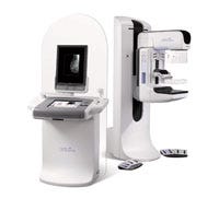
Designed to detect the presence of calcifications, masses, distortions, and asymmetries in breast tissue that could indicate breast cancer, mammograms can be both physically and mentally uncomfortable for many patients. While feeling stressed in such an environment is understandable, stress can affect results. And receiving clear, accurate results can make a difference in the treatment options and success rates for patients diagnosed with breast cancer. Aiming to optimize patient comfort to yield better results, Hologic (Bedford, MA) spent nine years developing the first commercially available breast cancer screening and diagnostic technology to deliver on the promise of breast tomosynthesis.
|
After the company's Selenia 2-D digital mammography system gained premarket approval in 2002, Hologic turned its attention to developing a 3-D version. "There's been a lot of work on 3-D breast tomosynthesis for years, but computers were too slow, and digital detectors were too slow," explains Georgia Hitzke, vice president, clinical services and product management for Hologic. "It was the right time because the detectors and computers were able to do this in a screening environment."
In a 2-D mammography image, objects of interest can be hidden in overlapping tissue, while normal breast structures that overlap may appear abnormal. Tomosynthesis is a 3-D imaging mode that reduces or eliminates this tissue superimposition effect, however. In Hologic's Selenia Dimensions tomosynthesis 3-D system, the breast is kept stationary between the detector housing and a compression paddle. The x-ray tube moves in an arc, taking a series of low-dose images from different angles. The slices are then reconstructed by a computer and transmitted to the diagnostic workstation for review by a radiologist. "We can take a 2-D and 3-D image under one compression," Hitzke says. "We can do both images at the same time."
To help incorporate the 3-D functionality with ergonomic components, Hologic sought a design partner and paired with Farm Design Inc. (Hollis, NH), a product development company that focuses on medical device design. Over the course of a year, Farm Design aided in the development of the Selenia Dimensions by creating an entirely new work station and full redesign of the digital detector system where the patient is postioned. Included in this work was human factors engineering, studying how patients interact with the device to maximize comfort and creating a unique graphical touch screen interface and proprietary x-ray exposure controls for operators to eliminate repetitive joint motion and user fatigue.
The design approach, according to Darrin Manke, director and program manager at Farm Design, was to make everything flush. This meant creating a constant level surface so that objects do not stick out when operators or patients interact with the device. On the workstation portion of the Selenia Dimensions 3-D system, improvements were made to the user interface. The x-ray image display, for example, can rotate out in the field so that operators can interact with the patient while they are also looking at the screen.
Farm Design also created a new control method to activate the x-ray system. "Typically, there are two switches on the top of a workstation, and you have to press them simultaneously to operate [the system]. They are tough to push, so they can't be triggered accidentally. Operators' fingers and thumbs were getting very sore," Manke says. "We developed recessed handle areas, with handles you reach in to pull levers, much like a bicycle break. Using all four fingers allows the operators to be relaxed."
To better understand the patient's perspective, on the other hand, Manke and another member of his design team experienced mammograms firsthand. "We wanted to understand the entire process, including the compression, and how the patient is positioned," Manke explains. "It is very uncomfortable. Your head is one way, [your] arm another, and you're hugging the device."
Such discomfort during the screening process can impact results, Manke adds. "You want the patient relaxed so that you can pull more breast tissue into the x-ray field," he says. "Trying to maximize the comfort reduced the stress to get better imagery."
One of the most challenging design aspects of the 3-D system, however, involved positioning the patient's head out of the x-ray field. Because it is performing 3-D imaging, the tube head rotates during the imaging process. Thus, the patient's head must be kept out of the rotational field using a face shield. The Farm team had to determine if patients were pressing against the shield for comfort and were able to reduce the size of the shield without decreasing functionality. "There's always a balance in addressing human factors and ergonomics and developing that around advanced technology," Manke comments. "There are certain things we could have done that could have made patients more comfortable, but it would have sacrificed imaging quality."
In addition to Farm Design's contributions, the Hologic team had to design the x-ray tubes from the ground up, maximizing what they could to get the exam done quickly. Hitzke of Hologic notes that short compression time was required to avoid patient motion, which results in blurry images. Hologic also had to design the electronics and software to reconstruct the 3-D images.
Because the 3-D system generates many more images for a radiologist to review than 2-D systems, a CAD system is currently being tested in international clinical trials to help improve the usefulness of the tomosynthesis technique. Once this software is approved in the United States, it will help radiologists with diagnosis, increasing their confidence to rule out breast cancer without having to recall the patient for further examination.
"The potential is really great," Hitzke says. "It's amazing, even better than we all thought. We tried to make the image quality the best that it could be. We continue to find new ways to find cancer."
About the Author(s)
You May Also Like


