Originally Published in MDDI May 2001
May 1, 2001
Originally Published in MDDI May 2001
R&D HORIZONS
Advanced surgical techniques are becoming less invasive and less traumatic to patients, but navigating the body's complex tissues and organs requires innovative systems for guidance and control.
William Loob
Technological advances have enabled the use of minimally invasive surgery (MIS) in a broad range of surgical specialties in recent years. From early applications, such as arthroscopic meniscectomy and laparoscopic cholecystectomy, MIS techniques are being adapted to treatment of aneurysms, various cardiac procedures, and spinal procedures, among others. Improvements in image guidance, fiber-optics, and robotic control systems have enabled surgeons to exercise more precise movements in manipulating instruments in the patient. Widespread acceptance of MIS techniques, however, will require additional technology development in a range of fields. With patient demand driving healthcare providers toward the use of less-invasive procedures, device manufacturers will need to increase their capabilities in the areas of imaging, micromachining, robotic equipment and components, and software systems.
MAPPING THE PATIENT
|
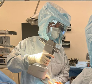
Intelligent OR systems are providing a range of new capabilities to surgeons performing MIS procedures. |
One of the technical capabilities required for navigating the MIS environment is a highly accurate and fast imaging modality to provide a detailed map of a patient's organs and tissues. Such maps enable surgeons to precisely position and manipulate instrumentation within the patient's body. Aaron Fenster, PhD, director of the Imaging Research Laboratories at the Robarts Research Institute (London, ON, Canada), oversees a variety of projects intended to address the limitations of current imaging technology for the MIS environment. It is instructive for researchers working on imaging technology associated with MIS to break up procedures into specific phases or modes of operation. These include treatment planning, instrument navigation through the patient, visualizing the site for performing the procedure, and postoperative monitoring. Each phase, Fenster says, makes distinct demands on imaging technology.
In part because of the imaging technology involved in MIS, virtual reality (VR) schemes have become a significant area of development. VR technology opens up the possibility of trying various scenarios on a virtual patient before the actual procedure is performed; however, it also makes heavy demands on technical capabilities. "For planning the procedure, we need user-friendly software that can display a full model of the patient for practicing the procedure in a virtual space," Fenster says. For such a system to operate effectively, haptic feedback devices are needed to re-create the feel of the instruments in actual use. That presents big difficulties in the functional modeling in the virtual patient, he continues. "An unexpected situation of intraoperative bleeding—now how do you simulate that?
"In guiding the instruments to the treatment site and performing the procedure, we need technologies that locate the instrument and image its local environment to be accurate and precise." But which imaging technology will prove to be most effective? Magnetic resonance imaging systems are sophisticated but are generally too cumbersome for the normally confined spaces of operating rooms, Fenster says. He believes that 3-D ultrasound systems will be the solution for intraoperative imaging because of the modality's compactness and real-time capabilities. On the other hand, he explains that the technology still needs improvement in some areas—particularly in image quality. "The challenges for 3-D ultrasound are improving the high-quality 3-D images and coming up with automated image interpretation algorithms. The radiologist is there in OR and the environment is already crowded and complicated." Tuning the imaging technology to temperature to help image the tissues and improving automated image-interpretation software might help surgeons navigate through a patient more accurately and provide more precise instrument control.
Other researchers working with advanced imaging systems agree that real-time imaging is needed to provide greater control of the surgical field within the patient. Frank Culicchia, MD, a New Orleans neurosurgeon, has been involved with the VectorVision system being developed by BrainLab (Munich, Germany). The system is a VR-based guidance system for surgeons working with complicated procedures. Culicchia says the system allows him to make smaller incisions and better localize tumors for more efficient and thorough tissue removal, but adds that the technology still works best in the pretreatment phase.
|
Robot-contolled MIS instruments provide a number of advantages. |
The VectorVision system compiles CT or MR images of the patient's brain into a 3-D model, which the surgeon uses to work out appropriate surgical approaches. With the current state of the technology, the image of the patient's brain only updates during the procedure to show an instrument's position. The present technology does not account for less-predictable changes, such as swelling or shrinking of tissues after instruments start cutting. "Under certain circumstances," Culicchia says, "the brain can shift after the initial steps in a procedure so that the image no longer corresponds exactly to the local position of the structures." For such conditions, Culicchia must still rely on tactile clues and on his skill and experience as a surgeon to compensate for changes in the brain tissues compared with the image displayed on the monitor. Although BrainLab is working on improvements to the system to accommodate the more advanced needs of such real-life working conditions, the problems are still viewed as difficult to overcome.
Fenster believes that the underlying technology needs for imaging boil down to a fundamental factor—real-time processing. "When I talk to colleagues in this field the standard phrase is 'speed, speed, speed and cost.'" Clearly the capabilities are desirable, he says, but the challenge is integrating all the necessary technologies into a functional and cost-effective system.
TAPPING INTO SPACE RESEARCH
Acquiring financing for new technology research and development is always a challenge. The cost of bringing innovative devices to market must be offset by the potential to recover that cost in an increasingly tight healthcare reimbursement environment. Some technologies, however, can get a helpful boost from high-tech fields outside medicine. Government and foundation grants for the development of robotics technology for space exploration has funded a number of projects that are aimed at improving or extending applications related to MIS.
A number of projects under way at the German Aerospace Center Institute for Robotics and Mechatronics (Wessling, Germany) are focused on development of a complete range of technologies that will improve the state of MIS applications. Much of the research has the general goal of yielding new systems and increasing understanding of control rules for micromechanisms that can be used in a variety of environments, including space. The groups at work on MIS applications, however, are finding that the requirements of confined surgical environments serve well as proving grounds for the development of delicate remotely operated mechanisms.
|
The Phantom haptic interface provides full force and torque feedback in the German Aerospace Center's MIS system. |
Ulrich Hagn, medical device development engineer in the German Aerospace Center's MIS project group, has designed remotely operated forceps, clamps, and suturing mechanisms. These devices are being integrated with robotic controls, imaging systems, and haptic interfaces to create an MIS environment based on principles of human-compliant motion. The researchers intend to create instrumentation that will provide surgeons with full sensory feedback, which will address the need for an intuitive basis for honing the surgeon's skills with the equipment.
The project team has produced a VR training device that enables a surgeon to become accustomed to how a haptic device responds to a corresponding image of a virtual organ. Others in the group are working on robotic controls that offer up to eight degrees of freedom to better model the dexterity of the human hand in operating the small surgical instruments. Although the project group has yet to find commercial partners to market the products, the researchers are working closely with clinicians who can demonstrate the systems.
Gleaning technology spin-offs from aerospace research is not a new practice, and some current MIS technologies come from fundamental work in space exploration. The demands of space research often mirror the technical features desired in MIS applications. One robotic system for assisting with MIS procedures is the AESOP endoscope positioner, developed by Computer Motion (Santa Barbara, CA). The robotic arm is designed to position and hold the endoscope steady during MIS procedures. The system extends the time during which surgeons can perform complex procedures because it assumes a function that human assistants cannot perform well over extended periods. NASA funded the robotics technology development so it might eventually be used in repairing satellites in space and inspecting payloads.
Computer Motion has continued its development programs from the robotics technology used in its AESOP system to integrate voice-command features that can facilitate control of the instrument. In addition, the firm developed a system called Zeus for endoscopic thoracic procedures and has obtained a CE mark for marketing the system in Europe. The company designed the system to be capable of performing heart bypass surgery on a beating heart.
PRODUCTS FOR SPECIFIC NEEDS
Although large-scale, research-intensive technologies for MIS applications often command most of the attention of the medical community, a significant part of new product development is being derived from efforts to address issues of MIS procedures performed on a more day-to-day basis. While the technology itself may not be necessarily critical to performing MIS, the products do fulfill important needs of physicians and equipment operators.
Surface-coatings manufacturer SurModics (Eden Prairie, MN) specializes in materials modifications that address lubricity, hemocompatibility, and infection resistance for medical devices. Last year, the firm developed a lubricity-enhancing coating for stainless-steel and nitinol guidewires used with arterial and venous catheters. SurModics developed its coating technology to allow device manufacturers to meet more stringent specifications for static friction. A lower static friction ratio can increase the responsiveness of guidewires to a surgeon's touch in starting a wire moving again after stopping momentarily or can offer greater torque control. The firm claims that its coating provides half the static friction ratio as conventional coating technologies.
Mectra Labs (Bloomington, IN) develops a variety of products for use in MIS procedures. The firm's MIS instruments are intended to combine multiple functions into a single device. Mectra's Nibblit device was designed to provide single-hand control when used in adhesiolysis, morcellation, or biopsy specimen collection without having to change between more-specialized instruments. The device was also designed for connection to irrigation and suction machines from different manufacturers, which allows hospitals to integrate the unit with existing equipment. Another of the company's products addresses a less technically complex but often overlooked problem of MIS—combating unpredictable fogging of endoscopes during MIS procedures. Mectra's Mr. Clear Anti-Fog Solution is applied directly to the endoscope lens prior to surgery.
Equipment testing is another arena for potential product development. The biomedical software specialist Premise Development Corp. (Avon, CT) focuses its efforts on the information technology that supports MIS instrumentation. The EndoTester is designed for calibrating and assessing the optical performance of endoscopes in the clinical setting. The firm's principals note that surgeons might sometimes change endoscopes several times during a procedure because of calibration loss or similar performance concerns. The device conducts several tests to verify specific modes of endoscope operation and was designed to operate with plug-in cards and standard computers used in clinical settings. Test functions include assessment of relative light loss, reflective symmetry, geometric distortion, modulation transfer function, and depth of focus.
One intended application of the tester applies to a new generation of multiuse, disposable endoscopes. The company notes that objective measures are needed to help clinicians determine the parameters for deciding end-of-effective-life criteria for multiuse scopes. Although manufacturers of such scopes often indicate on the labeling that product life is between 20 and 30 procedures, determining maximum useful life can be difficult. Testing optical performance will help set the end point, according to the firm.
EDUCATING SURGEONS
Joseph Adam, Premise Development CEO and cofounder, observes that equipment-testing products also enhance physicians' trust in new technologies. Instruments developed for MIS can challenge surgeons by changing the way they work as well as the way they receive information about the procedures they perform. Education and training are additional ways to gain clinicians' trust.
Most companies involved in developing sophisticated MIS-assistive systems for imaging and communication see great potential for physician education applications. Jonathan Silverstein, MD, assistant professor of surgery at the University of Illinois Health Sciences Center (Chicago) thinks that the heavy use of information technology in modern laparoscopic surgery lends itself well to teaching applications. As codirector of the university's Virtual Reality in Medicine Laboratory, Silverstein also is involved in the development of wearable VR displays. Distributed computing platforms and real-time imaging can connect an experienced surgeon to students in teaching procedures. Such systems will ultimately be used to help train physicians who will accept the technology as the normal environment for surgery.
Highly networked systems operating within information-intensive environments rely on advanced computing power. This presents a challenge to the current capabilities of affordable information systems available to hospitals. Fenster at Robarts Institute acknowledges that computing technology places limitations on VR technology associated with the high end of MIS system research—the application most useful in physician education and in presurgical treatment planning. "I like to compare it with simulators for training fighter pilots: They simulate very complex operating environments. I argue the surgical procedure is much more difficult and complex to simulate—but it is possible."
CONCLUSION
The scope of MIS procedures is expected to expand as instrumentation and guidance systems are refined. Yet the technology must still overcome a number of challenges. Reimbursement for new procedures will remain a key issue. In addition, the transfer of new procedural technologies into clinical practice will require improved methods for training practicing physicians.
William Loob is a medical writer living in Brooklyn, NY.
Photo courtesy of University Of Pittsburgh Medical Center
Photo courtesy of German Aerospace Center
Photo courtesy of Sensable Technologies
Copyright ©2001 Medical Device & Diagnostic Industry
About the Author(s)
You May Also Like


