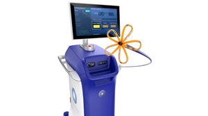
An infrared scanning system developed at Johns Hopkins University could detect melanoma.
April 1, 2010
In the battle against skin cancer, researchers are hoping that an infrared scanning device brings them one step closer to noninvasive, early disease detection. Developed at Johns Hopkins University, the device uses thermal imaging to detect the difference in temperature between healthy and cancerous tissue in pigmented skin.
“Our goal is to give an objective measurement as to whether a lesion is malignant,” says Rhoda Alani, professor at the Johns Hopkins Kimmel Cancer Center. “It could take much of the guesswork out of screening patients for skin cancer.” The researchers are working toward developing a device that diagnoses early-stage melanoma before the disease has a chance to spread.
Alani worked on the project with Cila Herman, professor of mechanical engineering at Johns Hopkins’ Whiting School of Engineering, and Muge Pirtini, a mechanical engineering doctoral student at the university. The National Science Foundation awarded Herman a $300,000 grant to develop a method to detect differences in heat below the skin’s surface.
|
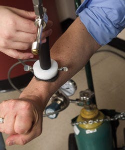
Before the targeted area of skin is scanned, it is cooled with a brief burst of compressed air. |
The researchers began by using a sensitive infrared camera. To change the skin’s temperature, they applied a burst of compressed air to the skin for one minute. This cooling of the skin isn’t harmful to patients. As the temperature of the skin returns to normal, researchers record infrared images for two to three minutes. Herman likened viewing the infrared images to using night vision goggles. Cancer cells usually reheat faster than healthy tissue, and the imaging can record any temperature differences between the tissues.
A 50-patient pilot study is examining the device’s sensitivity in detecting melanomas and precancerous lesions. Lesions are tested with the thermal imaging scanner, and a biopsy evaluates the presence of melanoma.
At this point, the researchers must further refine the scanner and its software, and develop diagnostic criteria for the malignant lesions, says Herman. Ultimately, they would like the final device to look like a handheld scanner that has the flexibility to be integrated into a full-body scanner for patients with several pigmented lesions.
About the Author(s)
You May Also Like


