The company hopes to accelerate stroke diagnosis and treatment for better patient outcomes.
July 12, 2021
Timing is critical when it comes to diagnosing and treating stoke patients. “If you get a stroke, your outcome is closely tied to how fast you get treatment,” Dr. Atul Gupta, chief medical officer for image-guided therapy at Royal Philips, told MD+DI. “You lose about 2 million neurons or brain cells every minute a stroke is in progress. That is why doctors have an expression: we say, ‘Time is Brain.’
“Every 30 minutes’ delay before treatment reduces the chance of a good outcome by 14%. Physicians in the emergency stroke setting are under immense pressure to rapidly reach a precise diagnosis so that clinically eligible stroke patients can benefit from a minimally invasive image-guided procedure called thrombectomy. The clock is ticking and if we can intervene and remove that tiny clot causing stroke quickly enough, disability can be minimized or even eliminated,” Gupta continued.
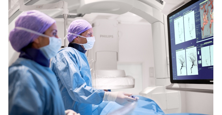
Philips hopes to make a difference in such timing with two recent developments regarding the diagnosis and treatment of stroke. First, the first patient has been enrolled in the WE-TRUST (Workflow optimization to rEduce Time to endovascular Reperfusion for Ultra-fast Stroke Treatment) trial study at Vall d’Hebron University Hospital, Barcelona, Spain, Philips reported July 7. This trial will study whether Philips’s Direct to Angio Suite workflow coupled with Philips Image Guided Therapy System, Azurion (pictured above), can improve outcomes for early time-window stroke patients (less than six hours after stroke onset).
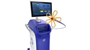
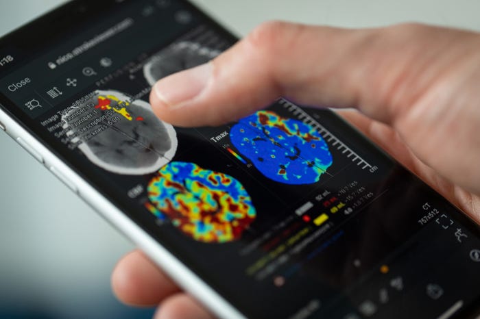
Philips also announced a strategic partnership agreement with NICO.LAB for use of a cloud-based, end-to-end artificial intelligence (AI) based stroke triage and management solution called StrokeViewer (shown below). The solution analyzes CT scan data with AI and automatically detects large vessel occlusion (LVO) and its location.
“Stroke teams currently lose valuable time due to hurdles in the workflow,” Gupta told MD+DI. “Our innovations are aimed at removing these hurdles across the full stroke care path, giving care teams the information and tools to make decisive decisions faster and accelerate stroke diagnosis and treatment so that patients have the best chance of complete recovery.”
Gupta says that through the WE-TRUST study, Philips is “pioneering a new approach bringing patients straight to the Philips Azurion interventional suite, diagnosing and treating them in the same room, with the aim to save valuable time to treatment.”
The approach involves use of the Direct to Angio Suite workflow, which uses improved cone-beam computed tomography (CBCT) to improve image quality and facilitate triage of patients. It reconstructs the stroke CBCT images using specially designed algorithms and filters to reduce artifacts caused by patient motion, cone beam hardening, and metal objects, all with the aim to improve the brain scan quality and support in clinical decision-making, Philips explained in a news release.
“In a single center randomized clinical trial in our center, the Direct to Angio Suite workflow has shown a significant improvement in clinical outcomes in patients who suffered a stroke,” said Dr. Marc Ribó, WE-TRUST co-Principal Investigator, Interventional Neurologist at the Vall d’Hebron University Hospital and researcher at the Stroke Research group at the Vall d’Hebron Research Institute (VHIR), stated in the release. “With the updated Stroke CBCT investigational device we can better facilitate triage of patients through improved image quality.”
And regarding Philips’s work with NICO.LAB, Gupta said the partnership “adds AI and cloud capabilities to our portfolio of stroke solutions to support decision making and connect information across the stroke care pathway, further enabling care teams to work quickly and act decisively.” Such AI-based CT scan analysis is shared with physicians at both primary stroke centers (general radiologists and neurologist) and intervention centers (stroke neurologists, interventional neurologists, and radiologists).
About the Author(s)
You May Also Like

