Sign up for the QMED & MD+DI Daily newsletter.
Nancy Crotti
January 18, 2017
3 Min Read
.svg?width=850&auto=webp&quality=95&format=jpg&disable=upscale "How Philips is Using Augmented Reality to Improve the Accuracy of Spinal Surgery")
In a preclinical study, neurosurgeons using augmented reality were able to more precisely place screws in cadavers' spines.
Nancy Crotti
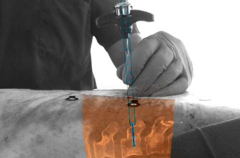
A preclinical study showed that Philips's augmented reality technology
helped increase the accuracy of pedicle screw placement in the
thoracic spines of cadavers versus freehand placement.
Royal Philips is working on an augmented-reality surgical navigation technology designed to help surgeons perform image-guided open and minimally-invasive spine surgery.
The technology has implications for thoracic spine fusion surgery in adults, pediatric spine surgery, and for cranial and trauma surgeries, according to a company statement. Located in the mid- to upper back, the thoracic spine is made up of smaller vertebrae than the lower, lumbar spine, making thoracic fusion surgery more challenging and dangerous to surrounding tissue and nerves.
A preclinical study of the new technology involving the placement of pedicle screws in the thoracic spines of cadavers demonstrated an 85% accuracy rate, versus 64% accuracy for freehand placement. Philips, Karolinska University Hospital (Stockholm, Sweden) and the Cincinnati Children's Hospital Medical Center collaborated on the study, published in the journal Spine.
Such improved accuracy implies lower complication rates, according to according to Ronald Tabaksblat, business leader of image-guided therapy systems at the Amsterdam-based company.
Surgical applications for virtual and augmented reality have been gaining attention since the release of Google Glass in 2013. (Philips Healthcare was an early proponent of that ill-fated technology, which was withdrawn in January 2015.) Using the low-cost VR system Google Cardboard, a London surgeon and and co-founder of a virtual and augmented reality company livestreameda procedure in which he removed a tumor from a patient's colon in April 2016.
Philips would need regulatory clearance to add its latest technology to its existing low-dose X-ray systems for clinical trials scheduled at Karolinska, Cincinnati Children's and about eight other sites globally, Tabaksblat said in an interview. If fully approved by FDA and the EU, Philips could deploy it to thousand of hospitals around the world, he noted.
The technology uses high-resolution optical cameras mounted on a flat panel X-ray detector to image the surface of the patient. By combining the external camera view and the internal 3D view of the patient acquired by the X-ray system, the technology constructs a 3D augmented-reality view of the external and internal anatomy. This real-time, 3D view of the patient's spine is designed to improve procedure planning, surgical tool navigation, and implant accuracy, and to reduce procedure time.
The technology could prove especially helpful in cranial surgery, because the pressure released by opening the cranium allows the brain to change shape. That movement can make presurgical images from MRI and CT scans less valuable to neurosurgeons, and make real-time imaging more valuable, according to Tabaksblat.
"There are significant challenges in that area, and we feel our technology can really make a difference there as well," he said. "We are starting with spine. We have a lot of experience in designing image guidance systems. You need to tailor them to the specific procedure."
Tabaksblat declined to discuss a timeline for a system to assist in brain surgery, but said fine-tuning the system for trauma surgeries would follow.
"We will really have to narrow it down to specific scenarios, but our customer are indicating that trauma is very interesting to look at," he said.
Nancy Crotti is a freelance contributor to Qmed.
About the Author(s)
You May Also Like