October 25, 2016
Quickly fabricated and customized, it may eliminate the need for animal testing.
Nancy Crotti
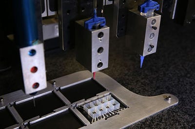
 Harvard University researchers have made what they are touting as the first entirely 3D-printed organ-on-a-chip with integrated sensing. By using new printable inks for multi-material 3-D printing, they were able to automate the fabrication process while increasing the complexity of the devices.
This new approach to manufacturing addresses a couple of the major problems with current organ-on-a-chip fabrication: it's labor-intensive and expensive. It may one day allow researchers to rapidly design organs-on-chips, also known as microphysiological systems, that match the properties of a specific disease or even an individual patient's cells, according to a university statement. The research is published in Nature Materials.
"This new programmable approach to building organs-on-chips not only allows us to easily change and customize the design of the system by integrating sensing but also drastically simplifies data acquisition," said Johan Ulrik Lind, first author of the paper and postdoctoral fellow at the Harvard John A. Paulson School of Engineering and Applied Sciences. Lind is also a researcher at Harvard's Wyss Institute for Biologically Inspired Engineering.
"Our microfabrication approach opens new avenues for in-vitro tissue engineering, toxicology, and drug-screening research," added Wyss faculty member Kit Parker, study co-author and a professor of bioengineering and applied physics at the Paulson School. Parker has also devised a "living" stingray robot in his quest to construct a human heart.
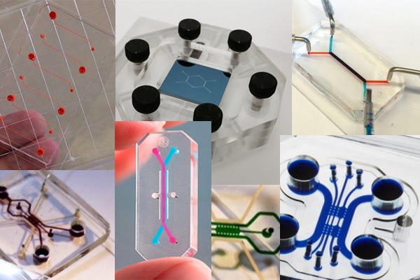
In the past year, researchers have produced a variety of body parts and even laboratories on chips. Organs-on-chips mimic the structure and function of native tissue, and have emerged as a promising alternative to traditional animal testing. Harvard researchers have developed microphysiological systems that mimic the microarchitecture and functions of lungs, hearts, tongues, and intestines.
The researchers on the latest study developed six different inks that integrated soft strain sensors within the micro-architecture of the tissue. In a single, continuous procedure, they 3-D printed those materials into a cardiac microphysiological device with integrated sensors.
The chip contains multiple wells, each with separate tissues and integrated sensors, allowing researchers to study many engineered cardiac tissues at once. To demonstrate the efficacy of the device, the team performed drug studies and longer-term studies of gradual changes in the contractile stress of engineered cardiac tissues, which can occur over the course of several weeks.
"Researchers are often left working in the dark when it comes to gradual changes that occur during cardiac tissue development and maturation because there has been a lack of easy, non-invasive ways to measure the tissue functional performance," said Lind. "These integrated sensors allow researchers to continuously collect data while tissues mature and improve their contractility. Similarly, they will enable studies of gradual effects of chronic exposure to toxins."
Nancy Crotti is a contributor to Qmed.
Like what you're reading? Subscribe to our daily e-newsletter.
[Image courtesy of Harvard University]
About the Author(s)
You May Also Like


