August 4, 2016
The reanimation allows for visualization of the unique functional anatomies within each heart, explains the University of Minnesota's Paul Iaizzo.
Chris Newmarker
|
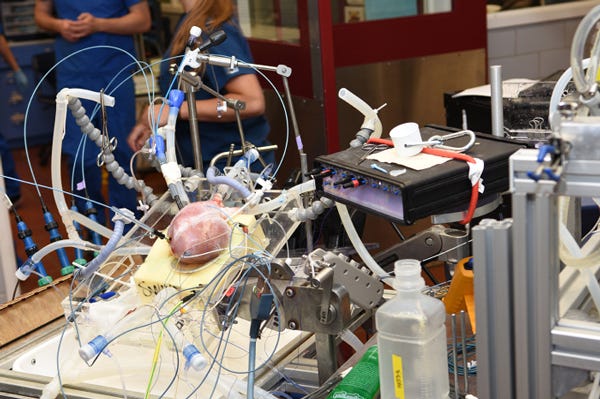
This living pig heart is hooked up on equipment to keep it beating at the University of Minnesota's Visible Heart Lab. Such equipment has also been used to reanimate 70 human hearts over the past 17 years. (Image courtesy of Visible Heart Lab) |
Watch enough Frankenstein movies, and you'll probably get excited about the idea of reanimated hearts. But such reanimation is much more than cool: it provides valuable insights for medical device designers, explains Paul Iaizzo, PhD, principal investigator at the University of Minnesota's Visible Heart Lab.
Since 1999, the Visible Heart Lab has reanimated 70 human hearts donated for research. Studying and visually recording cardiac device placement and performance within various reanimated human hearts enables visualization of the unique functional anatomies within each heart. "We can also perform comparative imaging studies employing multiple imaging modalities: e.g., videoscopes, fiber scopes, high speed cameras, echcardiography, and/or fluoroscopy," Iaizzo says.
Iaizzo shared a paper he has written that, among other things, further elaborates on the benefits. They include:
Giving device designers a way to rapidly and critically assess prototypes, expediting decisions around design;
Providing a way for clinicians to directly visualize how new or problematic clinical systems might be deployed in the heart;
Allowing patients and students at all levels to better educate themselves.
Iaizzo also noted that cardiac devices' future involves beating heart procedures such as transcatheter-delivered valves, repairs, closure systems, and pacemakers. Such procedures will likely utilize simultaneous multiple imaging modalities--methods the Visible Heart Lab is already using with reanimated hearts.
The lab has a free online Atlas of Human Cardiac Anatomy with more than 5000 image and movies. It's also posted some footage from reanimated human hearts on YouTube.
Here's a mechanical aortic valve inside a reanimated heart:
And this is the right ventricular inflow tract:
This is a mitral valve:
Here's a tricuspid valve:
And this is a right ventricular outflow tract:
Chris Newmarker is senior editor of Qmed. Follow him on Twitter at @newmarker.
Like what you're reading? Subscribe to our daily e-newsletter.
About the Author(s)
You May Also Like


